 NEET Notes
Home > Biology > Zoology > Neural Control and Coordination
Neural Control and Coordination
On this page · 13 sections
- 01Syllabus map
- 02The neural system
- 03Human neural system
- 04Neuron: structure and types
- 05Generation and conduction of nerve impulse
- 06Transmission of impulses
- 07Brain: forebrain
- 08Brain: midbrain and hindbrain
- 09Removed topics: reflex action, eye, ear and spinal cord
- 10Table 1 - Types of neurons
- 11Table 2 - Divisions of the human neural system
- 12Table 3 - Parts of the brain and what each does
- 13Ladder - The confusions that cost marks
Neurons, nerve impulse, synapses and the brain, from NCERT Chapter 18. Reflex action and the eye and ear are out of the current syllabus and appear only as a short summary.
Syllabus map
- NMC Unit: BIO-U05, Human Physiology. NCERT Chapter 18, Neural Control and Coordination.
- Subtopics in the current NMC syllabus: neuron and nerves; nervous system in humans (central, peripheral and visceral nervous system); generation and conduction of nerve impulse.
- What this chapter covers: the neural system in Hydra, insects and vertebrates; the human neural system (CNS, PNS, somatic and autonomic divisions, visceral nervous system); the structure and types of neurons; resting potential, action potential and impulse conduction; electrical and chemical synapses; the forebrain, midbrain and hindbrain.
- Removed from both the rationalised NCERT Chapter 18 and the current NMC syllabus: reflex action, sense organs (the eye and the ear), and the spinal cord as a separate section. They appear in this chapter only as one short summary (section 9), for older PYQs and mock tests.
- Estimated revision time: 12 minutes. Jump links: Syllabus Map, Neural System, Human Neural System, Neuron, Nerve Impulse, Transmission, Forebrain, Midbrain and Hindbrain, Removed Topics, Tables, Trap Boxes, PYQs, Chapter Test.
The neural system
Coordination is the process through which two or more organs interact and complement the functions of one another. In our body, the neural system and the endocrine system jointly coordinate and integrate all the activities of the organs so that they function in a synchronised fashion.
The neural system provides an organised network of point-to-point connections for quick coordination. The endocrine system provides chemical integration through hormones.
The neural system of all animals is composed of highly specialised cells called neurons, which can detect, receive and transmit different kinds of stimuli.
- In Hydra, the neural system is a network of neurons: there is no brain and no central control.
- In insects, the neural system is better organised: a brain is present along with a number of ganglia and neural tissues.
- In vertebrates, the neural system is far more developed, with a distinct brain and spinal cord.
Human neural system
The human neural system is divided into two parts: the central neural system (CNS) and the peripheral neural system (PNS).
The CNS includes the brain and the spinal cord. It is the site of information processing and control.
The PNS comprises all the nerves of the body associated with the CNS. The nerve fibres of the PNS are of two types: afferent fibres, which transmit impulses from tissues and organs to the CNS, and efferent fibres, which transmit regulatory impulses from the CNS to the concerned peripheral tissues and organs.
Extra information (beyond NCERT): the PNS is also described by the nerves themselves: cranial nerves (12 pairs) and spinal nerves (31 pairs). NCERT does not give these numbers.
The PNS is divided into the somatic neural system and the autonomic neural system:
- The somatic neural system relays impulses from the CNS to the skeletal muscles. It is voluntary: you decide to move your arm.
- The autonomic neural system transmits impulses from the CNS to the involuntary organs and smooth muscles of the body, such as the heart and the gut. It is involuntary.
- The autonomic neural system is further classified into the sympathetic neural system and the parasympathetic neural system.
Extra information (beyond NCERT): the sympathetic division is often described as 'fight or flight' and the parasympathetic as 'rest and digest'. NCERT names the two divisions but does not use these phrases.
The visceral nervous system is the part of the peripheral nervous system that carries impulses from the CNS to the viscera and from the viscera to the CNS.
Practice1 question
Try it now. A wrong answer costs nothing here, try again until you get it.
Which of the following is controlled by the autonomic nervous system?
The autonomic nervous system controls involuntary actions such as the heartbeat, peristalsis and the secretion of glands. The somatic nervous system relays impulses from the CNS to the skeletal muscles, which is voluntary.
Neuron: structure and types
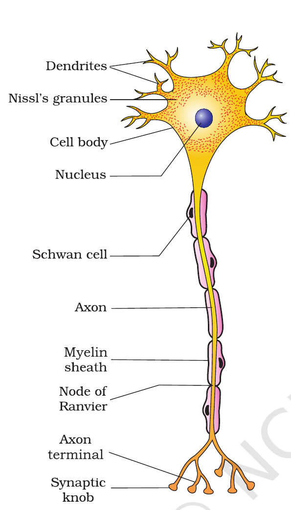
A neuron is the structural and functional unit of the neural system. It has three parts: the cell body, dendrites and the axon.
The cell body (cyton or soma) contains cytoplasm with typical cell organelles, a nucleus, and certain granular bodies called Nissl's granules. It is the part of the neuron that carries out the metabolic activities of the cell.
Dendrites are short fibres that project out of the cell body. They transmit impulses towards the cell body.
The axon is a long fibre, the distal end of which is branched. Each branch terminates as a bulb-like structure called the synaptic knob, which possesses synaptic vesicles containing chemicals called neurotransmitters.
The axon may be myelinated or non-myelinated. The myelinated nerve fibres are enveloped with Schwann cells, which form a myelin sheath around the axon. The gaps between two adjacent myelin sheaths are called nodes of Ranvier.
Extra information (beyond NCERT): in the central nervous system, myelin is made by cells called oligodendrocytes, not by Schwann cells. NCERT names only Schwann cells, but NEET asked this distinction in 2017.
Based on the number of axons and dendrites, neurons are of three types:
- Multipolar: one axon and two or more dendrites, e.g. neurons of the cerebral cortex.
- Bipolar: one axon and one dendrite, e.g. neurons of the retina of the eye.
- Unipolar: cell body with one axon only, found usually in the embryonic stage.
Generation and conduction of nerve impulse
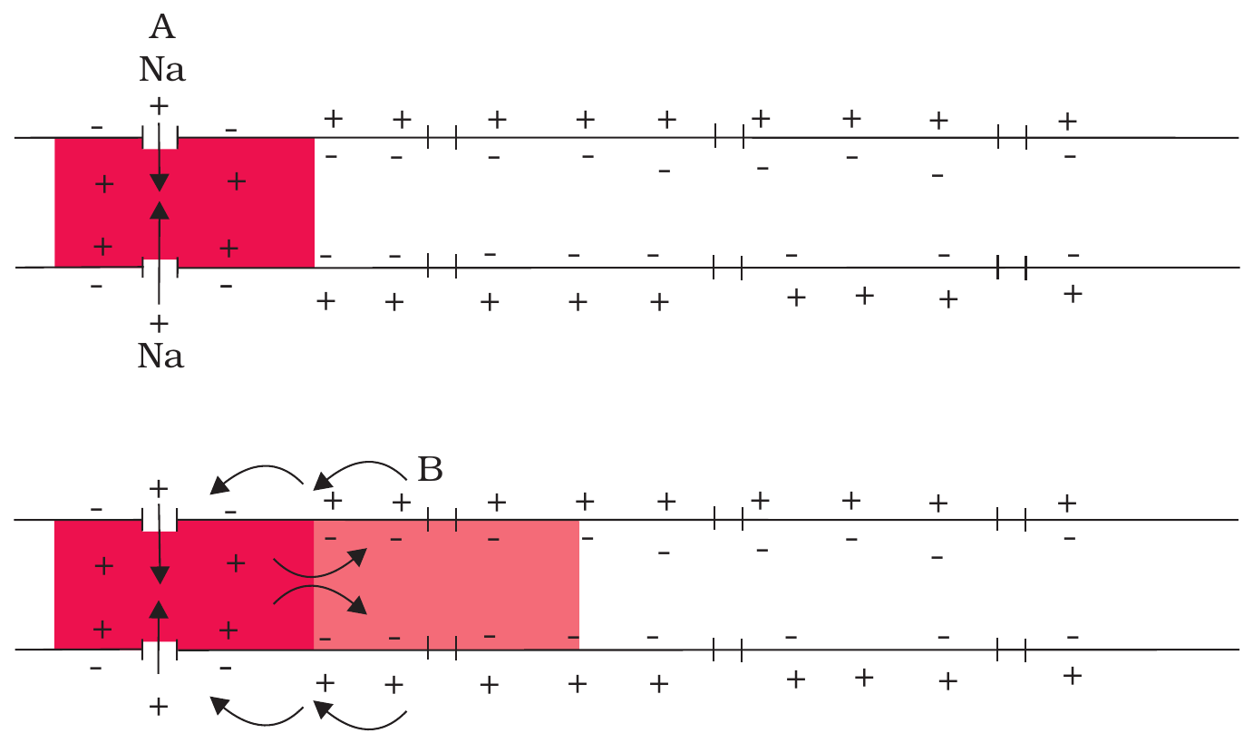
When a neuron is not conducting any impulse, the axonal membrane is in a resting state: the outer surface of the membrane is positively charged and the inner surface is negatively charged. The electrical potential difference across the resting plasma membrane is called the resting potential. Extra information (beyond NCERT): its value is about -70 mV (inside negative).
The ionic gradients across the resting membrane are maintained by the active transport of ions by the sodium-potassium pump, which transports 3 $Na^+$ outwards for 2 $K^+$ into the cell. As a result, the outer surface of the axonal membrane possesses a positive charge while its inner surface becomes negatively charged.
When a stimulus is applied at a site on the polarised membrane, the membrane at that site becomes freely permeable to $Na^+$. There is a rapid influx of $Na^+$ ions, and the inner side of the membrane becomes positively charged while the outer side becomes negative. This reversal of polarity is called depolarisation, and the membrane at the site is said to be in the excited state.
The electrical difference across the membrane at the site of excitation is called the action potential, which is in fact termed a nerve impulse.
At the site of excitation, the membrane then becomes permeable to $K^+$, so $K^+$ ions rush out. This restores the resting potential: the inner side becomes negative again. This is repolarisation. The membrane is then ready to receive the next stimulus.
The action potential is propagated along the axon from one point to the next. In myelinated fibres, the impulse jumps from one node of Ranvier to the next. It is much faster than conduction in non-myelinated fibres, where the impulse travels continuously along the whole length of the axon. Extra information (beyond NCERT): this jumping is called saltatory conduction.
Practice1 question
Try it now. A wrong answer costs nothing here, try again until you get it.
During depolarisation of a neuron, what happens?
When a stimulus is applied, the membrane becomes permeable to $Na^+$ and $Na^+$ rushes in, so the inside becomes positive: depolarisation. $K^+$ moves out later, during repolarisation.
Transmission of impulses
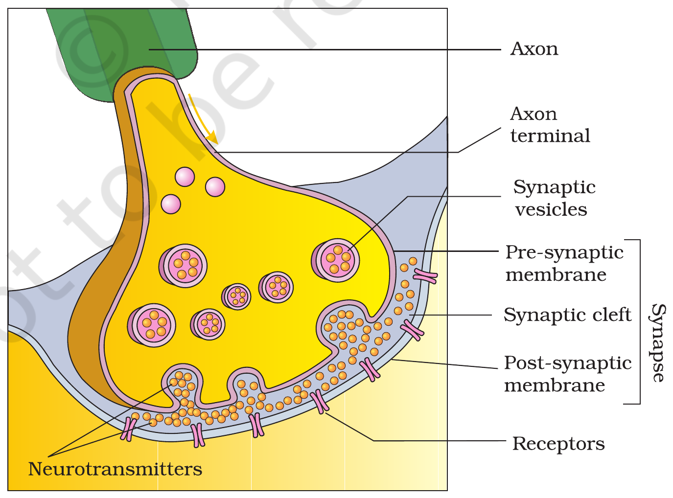
A nerve impulse is transmitted from one neuron to another through a junction called a synapse. A synapse is formed by the membranes of a pre-synaptic neuron and a post-synaptic neuron, which may or may not be separated by a gap called the synaptic cleft.
There are two types of synapses: electrical synapses and chemical synapses.
- Electrical synapses: the membranes of the pre-synaptic and post-synaptic neurons are in close proximity. The impulse is transmitted directly from one neuron to the other, so transmission is very fast. Electrical synapses are rare in our system.
- Chemical synapses: the membranes of the pre-synaptic and post-synaptic neurons are separated by a fluid-filled space called the synaptic cleft. When an impulse arrives at the axon terminal, the synaptic vesicles release neurotransmitters into the cleft. The neurotransmitter binds to receptors on the post-synaptic membrane, opens ion channels, and generates a new potential in the post-synaptic neuron.
Extra information (beyond NCERT Ch 18; from Ch 17, Sec 17.2.2): Acetylcholine is the best-known neurotransmitter. At the neuromuscular junction, acetylcholine released from the motor neuron binds to receptors on the muscle fibre and triggers contraction.
Most synapses in the human body are chemical synapses. The synaptic cleft means transmission is slower than at an electrical synapse, but the chemical step allows the signal to be strengthened, weakened or blocked.
Brain: forebrain
The brain is the central information processing organ of our body, and acts as the command and control system. It controls the voluntary movements, balance of the body, functioning of vital involuntary organs (e.g. lungs, heart, kidneys), thermoregulation, hunger and thirst, circadian (24-hour) rhythms, activities of several endocrine glands, and human behaviour. It is also the site for processing of vision, hearing, speech, memory, intelligence, emotions and thoughts.
The brain is protected by the skull. Inside the skull, the brain is covered by three layers called cranial meninges: the outer layer is called the dura mater, the middle layer is called the arachnoid, and the inner layer (which is in contact with the brain tissue) is called the pia mater.
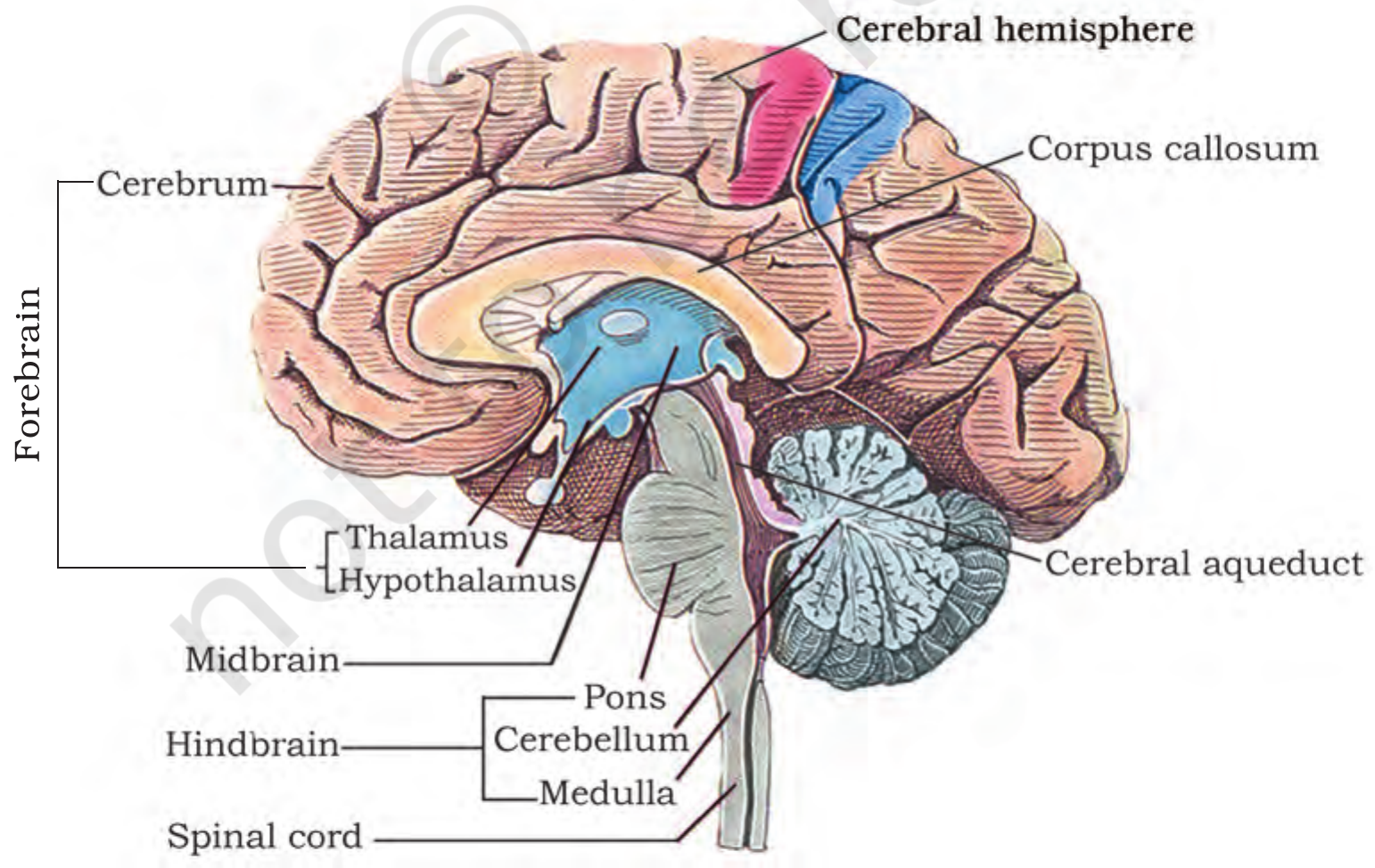
The brain can be divided into three major parts: (i) forebrain, (ii) midbrain, and (iii) hindbrain.
The forebrain consists of the cerebrum, thalamus and hypothalamus.
The cerebrum is the largest and most complex part of the brain. It is divided into two cerebral hemispheres by a deep cleft. The hemispheres are connected by a tract of nerve fibres called the corpus callosum. The outer part of the cerebrum is the cerebral cortex, made of grey matter; the inner part is made of white matter. The cerebrum is the seat of intelligence, memory and willpower: it controls voluntary movement, speech, smell, taste, hearing, vision and learning.
Extra information (beyond NCERT): The diencephalon is the part of the forebrain that encloses the thalamus and the hypothalamus. The thalamus is a major coordination centre for sensory and motor signalling: almost all sensory information passes through it on its way to the cortex. The cerebrum wraps around the thalamus.
The hypothalamus lies at the base of the thalamus. It contains a number of centres which control body temperature, urge for eating and drinking. It also contains several groups of neurosecretory cells, which secrete hormones called hypothalamic hormones. These hormones regulate the synthesis and secretion of pituitary hormones. Extra information (beyond NCERT): the pituitary is often called the master gland.
The cerebral cortex contains motor areas, sensory areas and large regions that are neither clearly sensory nor motor in function. These regions, called the association areas, are responsible for complex functions like intersensory associations, memory and communication.
The inner parts of the cerebral hemispheres and a group of associated deep structures like the amygdala and hippocampus form a complex structure called the limbic system, which is involved in the regulation of sexual behaviour, expression of emotional reactions (e.g. excitement, pleasure, rage and fear), and motivation. The sense of smell is also processed here.
Practice1 question
Try it now. A wrong answer costs nothing here, try again until you get it.
Which part of the brain controls body temperature?
The hypothalamus is the thermoregulatory centre: it controls body temperature, and also hunger, thirst and emotions. The thalamus is a major coordination centre for sensory and motor signalling.
Brain: midbrain and hindbrain
The midbrain is located between the thalamus/hypothalamus of the forebrain and pons of the hindbrain. A canal called the cerebral aqueduct passes through the midbrain. The dorsal portion of the midbrain consists mainly of four round swellings (lobes) called corpora quadrigemina. Extra information (beyond NCERT): the upper two are called the superior colliculi (visual reflexes) and the lower two the inferior colliculi (auditory reflexes).
The hindbrain comprises the pons, cerebellum and medulla (medulla oblongata).
The pons consists of fibre tracts that interconnect different regions of the brain.
The cerebellum has a very convoluted surface in order to provide the additional space for many more neurons. Extra information (beyond NCERT): the cerebellum coordinates body movements, maintains posture and balance, and makes voluntary movements smooth and precise.
The medulla is connected to the spinal cord. It contains centres that control respiration, cardiovascular reflexes and gastric secretions.
The midbrain, pons and medulla together form the brain stem: the stalk that connects the brain to the spinal cord and carries all the tracts running between them.
Removed topics: reflex action, eye, ear and spinal cord
Extra information (removed from both rationalised NCERT Ch 18 and the current NMC syllabus, for older PYQs / mock-test reference only): These topics were in older NCERT editions but are not in the current chapter or syllabus. Learn them only after the NCERT content above is solid.
- Reflex action: an involuntary, almost instantaneous response to a stimulus. It runs through a reflex arc: receptor, sensory (afferent) neuron, interneuron in the spinal cord, motor (efferent) neuron, effector.
- Eye: the photoreceptors are rods (dim light, rhodopsin) and cones (bright light, colour vision, iodopsin). The fovea has only cones and gives the sharpest vision; the blind spot has no photoreceptors.
- Ear: the organ of Corti sits on the basilar membrane inside the cochlea. Its hair cells convert sound vibrations into nerve impulses.
- Spinal cord: the long continuation of the medulla running down the vertebral column. Grey matter lies inside in an H-shape and white matter outside. It conducts impulses to and from the brain and is the centre for spinal reflex actions.
Practice1 question
Try it now. A wrong answer costs nothing here, try again until you get it.
Which of the following topics is NOT in the current NMC syllabus for Neural Control and Coordination?
Reflex action was removed from the rationalised NCERT Chapter 18 and is not listed in the current NMC syllabus. The other three topics are listed in the syllabus.
Table 1 - Types of neurons
| Type | Axons | Dendrites | Where it is found |
|---|---|---|---|
| Multipolar | One | Two or more | Cerebral cortex |
| Bipolar | One | One | Retina of the eye |
| Unipolar | One | None (cell body with one axon only) | Usually in the embryonic stage |
Source: NCERT XI, Ch 18, 18.3.
Table 2 - Divisions of the human neural system
| Division | Parts | Control | Example |
|---|---|---|---|
| Central neural system | Brain and spinal cord | Information processing and control | Thinking, memory |
| Somatic neural system | Cranial and spinal nerves to skeletal muscle | Voluntary | Moving an arm, walking |
| Sympathetic (autonomic) | Nerves to involuntary organs | Involuntary | Heart rate rises, pupils dilate, sweating |
| Parasympathetic (autonomic) | Nerves to involuntary organs | Involuntary | Heart rate falls, digestion increases |
| Visceral nervous system | Nerves between CNS and viscera | Both directions | Signals from gut to brain and back |
Extra information (beyond NCERT): the sympathetic division is often described as 'fight or flight' and the parasympathetic as 'rest and digest'.
Source: NCERT XI, Ch 18, 18.2.
Practice1 question
Try it now. A wrong answer costs nothing here, try again until you get it.
Which of the following is a part of the central neural system?
The brain and spinal cord form the central neural system. Cranial nerves, spinal nerves and the autonomic nerves are part of the peripheral neural system.
Table 3 - Parts of the brain and what each does
| Part | Division | Key function |
|---|---|---|
| Cerebrum | Forebrain | Intelligence, memory, willpower, voluntary movement, speech, vision, hearing |
| Thalamus | Forebrain | Coordination centre for sensory and motor signalling |
| Hypothalamus | Forebrain | Body temperature, hunger, thirst, emotions; controls the pituitary |
| Limbic system | Forebrain | Sexual behaviour, emotions, motivation, sense of smell |
| Midbrain | Midbrain | Corpora quadrigemina (four round swellings); receives and integrates visual, tactile and auditory inputs |
| Pons | Hindbrain | Fibre tracts interconnecting brain regions |
| Cerebellum | Hindbrain | Very convoluted surface, providing space for many more neurons |
| Medulla oblongata | Hindbrain | Respiration, cardiovascular reflexes, gastric secretions |
| Brain stem | Midbrain + pons + medulla | Connects brain to spinal cord; carries all tracts |
Extra information (beyond NCERT): the cerebellum coordinates body movements, maintains posture and balance, and makes voluntary movements smooth and precise. The upper two corpora quadrigemina are called the superior colliculi (visual reflexes) and the lower two the inferior colliculi (auditory reflexes).
Source: NCERT XI, Ch 18, Sec 18.4.1, 18.4.2 and 18.4.3.
Ladder - The confusions that cost marks
| Trap | Wrong | Right |
|---|---|---|
| Myelin maker | Schwann cells make myelin everywhere | Schwann cells in the PNS; oligodendrocytes in the CNS |
| Depolarisation | $K^+$ influx depolarises the neuron | $Na^+$ influx depolarises; $K^+$ efflux repolarises |
| Autonomic | The autonomic neural system is voluntary | The autonomic neural system is involuntary; the somatic neural system is voluntary |
| Brain stem | The brain stem is only the medulla | The midbrain, pons and medulla together form the brain stem |
| Brain seats | The cerebellum is the largest part of the brain | The cerebrum is the largest and most complex part of the brain |
| Grey matter | The cerebral cortex is white matter | The cerebral cortex is grey matter (neuron cell bodies are concentrated there); the inner part is white matter |
| Corpus callosum | The corpus callosum connects the two hemispheres of the cerebellum | The corpus callosum is a tract of nerve fibres connecting the two cerebral hemispheres |
| Synapse speed | Chemical synapses are faster than electrical ones | Electrical synapses are faster (direct); chemical synapses are slower but can be strengthened or blocked |
Source: NCERT XI, Ch 18; NEET 2017, 2018, 2019, 2022, 2024.
Trap Boxes
Schwann cells make myelin everywhere?
Wrong idea: Myelin is made by Schwann cells in the whole nervous system.
Myelin is made by Schwann cells in the peripheral nervous system and by oligodendrocytes in the central nervous system. NCERT names only Schwann cells, but NEET asked the distinction in 2017.
NCERT XI, Ch 18, 18.3
$K^+$ influx causes depolarisation?
Wrong idea: When a neuron depolarises, potassium ions rush into the cell.
Depolarisation is caused by a rapid influx of $Na^+$ ions. $K^+$ ions rush out later, during repolarisation, when the resting potential is restored.
NCERT XI, Ch 18, 18.3.1
Sympathetic is rest and digest?
Wrong idea: The sympathetic nervous system brings the body back to rest and digest.
Sympathetic prepares the body for action: heart rate rises, pupils dilate, sweating increases. Parasympathetic calms the body down: heart rate falls and digestion increases. Extra information (beyond NCERT): these are often called 'fight or flight' and 'rest and digest'.
NCERT XI, Ch 18, 18.2
The midbrain is part of the forebrain?
Wrong idea: The midbrain is one of the three parts of the forebrain.
The brain has three major parts: forebrain, midbrain and hindbrain. The midbrain lies between the thalamus/hypothalamus of the forebrain and the pons of the hindbrain.
NCERT XI, Ch 18, Sec 18.4.2
The cerebral cortex is white matter?
Wrong idea: The cerebral cortex is made of white matter.
The cerebral cortex is grey matter: neuron cell bodies are concentrated there, giving the grey colour. The inner part of the cerebral hemisphere is white matter.
NCERT XI, Ch 18, Sec 18.4.1
Chapter test
5 NEET PYQs. +4 for a correct answer, -1 for a wrong answer, exactly like NEET. Finish the test to earn a medal.
Q1: NEET (UG) 2024, Code R3, Q178
Match List I with List II:
| List I | List II |
|---|---|
| A. Pons | I. Provides additional space for Neurons, regulates posture and balance. |
| B. Hypothalamus | II. Controls respiration and gastric secretions. |
| C. Medulla | III. Connects different regions of the brain. |
| D. Cerebellum | IV. Neuro secretory cells. |
Pons connects different regions of the brain (III); hypothalamus has neurosecretory cells (IV); medulla controls respiration and gastric secretions (II); cerebellum provides additional space for neurons and regulates posture and balance (I).
Q2: NEET (UG) 2022, Code R1, Q189
Select the incorrect statement regarding synapses:
Transmission across a chemical synapse is slower than across an electrical synapse, because the chemical step takes time. Electrical synapses are rare, fast and direct; chemical synapses use neurotransmitters.
Q3: NEET (UG) 2019, Code S2, Q57
Which part of the brain is responsible for thermoregulation?
The hypothalamus contains centres that control body temperature, hunger and thirst. The medulla controls respiration and cardiovascular reflexes; the cerebrum is the seat of intelligence and memory; the corpus callosum connects the two cerebral hemispheres.
Q4: NEET (UG) 2018, Code AA, Q142
Which of the following structures or regions is incorrectly paired with its function?
The limbic system (amygdala, hippocampus) regulates emotion, sexual behaviour and motivation. The fibre tracts that interconnect different regions of the brain describe the pons, not the limbic system.
Q5: NEET (UG) 2017, Code A, Q171
Myelin sheath is produced by
Myelin sheath is produced by Schwann cells in the peripheral nervous system and by oligodendrocytes in the central nervous system. Astrocytes support neurons but do not make myelin; osteoclasts are bone cells.
Score: 0 · Answered 0 of 5
Flashcards
Tap a card to flip it over and see the answer. Tap it again to flip it back.
What is the resting potential?
Tap to flipThe electrical potential difference across the resting plasma membrane: the inside of the axon is negative relative to the outside. Extra information (beyond NCERT): the value is about -70 mV.
Tap to flip backPut the path of a nerve impulse through a neuron in order.
Tap to flipDendrite, cell body, axon, axon terminal.
Tap to flip backWhy do myelinated fibres conduct impulses faster than non-myelinated fibres?
Tap to flipIn myelinated fibres the impulse jumps from node to node, so it travels much faster than the continuous conduction along a non-myelinated fibre. Extra information (beyond NCERT): this jumping is called saltatory conduction.
Tap to flip backWhat connects the two cerebral hemispheres?
Tap to flipThe corpus callosum: a tract of nerve fibres connecting the two cerebral hemispheres.
Tap to flip backWhich cells make the myelin sheath in the PNS and in the CNS?
Tap to flipSchwann cells make myelin in the peripheral nervous system; oligodendrocytes make it in the central nervous system. NCERT names only Schwann cells.
Tap to flip backName the three cranial meninges from outside to inside.
Tap to flipDura mater (outer), arachnoid (middle), pia mater (inner).
Tap to flip backWhich fibres carry impulses towards the CNS, and which carry them away?
Tap to flipAfferent fibres carry impulses from tissues and organs to the CNS; efferent fibres carry regulatory impulses from the CNS to the tissues and organs.
Tap to flip backWhat are Nissl's granules?
Tap to flipGranular bodies in the cell body of a neuron, along with the usual cell organelles.
Tap to flip backWhat is the cerebral aqueduct?
Tap to flipThe canal that passes through the midbrain.
Tap to flip backWhat is a synapse?
Tap to flipA junction between two neurons where an impulse is transmitted from one neuron to another. The membranes of the pre- and post-synaptic neurons may or may not be separated by a gap called the synaptic cleft.
Tap to flip back