 NEET Notes
Home > Biology > Zoology > Locomotion and Movement
Locomotion and Movement
On this page · 17 sections
- 01Syllabus map
- 02Types of movement
- 03Muscle: three types
- 04Skeletal muscle fibre and the sarcomere
- 05Structure of contractile proteins
- 06Mechanism of muscle contraction
- 07Red and white muscle fibres
- 08Axial skeleton: skull and vertebral column
- 09Sternum and ribs
- 10Appendicular skeleton: pectoral girdle and arm
- 11Appendicular skeleton: pelvic girdle and leg
- 12Joints
- 13Disorders of muscular and skeletal system
- 14Table 1 - Three types of muscle
- 15Table 2 - Axial vs appendicular skeleton
- 16Table 3 - Joints at a glance
- 17Ladder - The confusions that cost marks
Muscle contraction, the skeleton, joints and disorders, straight from NCERT Chapter 17.
Syllabus map
- NMC Unit: BIO-U05, Human Physiology. NCERT Chapter 17, Locomotion and Movement.
- Subtopics in this chapter (NCERT XI, Ch 17): Types of movement. Muscle. Structure of contractile proteins. Mechanism of muscle contraction. Skeletal system. Joints. Disorders of muscular and skeletal system.
- Subtopic checklist: (1) amoeboid, ciliary and muscular movement (NCERT lists three types; flagellar movement of sperm is introduced in 17.2); (2) the three types of muscle; (3) structure of a skeletal muscle fibre and the sarcomere; (4) actin, myosin and the sliding filament mechanism; (5) red and white muscle fibres; (6) axial skeleton: skull, vertebral column, sternum and ribs; (7) appendicular skeleton: pectoral and pelvic girdles and limbs; (8) joints; (9) disorders of muscle and skeleton.
- Extra information (beyond NCERT): the slow and fast fibre labels, red fibres working without fatigue and white fibres in short bursts; the biceps example; fusiform (spindle-shaped) and uninucleate visceral fibres; cardiac muscle contracting rhythmically and not tiring easily; the patella as a sesamoid bone; the femur as the strongest bone; the tibia and fibula as the shank; the elbow as a hinge joint and the femur-acetabulum as a ball and socket joint; the sacral vertebra as 5 fused vertebrae and the coccygeal as 4 fused vertebrae.
- Excluded topics: neural control and coordination (Chapter 18) and chemical coordination and integration (Chapter 19). Do not import their facts here.
- Estimated revision time: 10 minutes. Jump links: Syllabus Map, Types of Movement, Muscle, Contractile Proteins, Contraction, Skeletal System, Joints, Disorders, Tables, Trap Boxes, PYQs, Chapter Test.
Types of movement
Movement is one of the significant features of living beings. Animals and plants exhibit a wide range of movements. Streaming of protoplasm in the unicellular organisms like Amoeba is a simple form of movement. Movement of cilia, flagella and tentacles are shown by many organisms.
Human beings can move limbs, jaws, eyelids, tongue, etc. Some of the movements result in a change of place or location. Such voluntary movements are called locomotion. Walking, running, climbing, flying and swimming are all forms of locomotion. All locomotions are movements but all movements are not locomotions.
Cells of the human body exhibit three main types of movements:
- Amoeboid movement: some specialised cells in our body like macrophages and leucocytes in blood exhibit amoeboid movement. It is effected by pseudopodia formed by the streaming of protoplasm.
- Ciliary movement: this takes place in most of our internal tubular organs which are lined by ciliated epithelium, for example the trachea and the oviducts.
- Muscular movement: movement of our limbs, jaws, tongue, etc., requires muscular movement. The contractile property of muscles is effectively used for locomotion and other movements by human beings and majority of multicellular organisms.
Flagellar movement is not one of the three types in NCERT 17.1. NCERT introduces it in 17.2: flagellar movement helps in the swimming of spermatozoa.
Muscle: three types
Muscles are specialised tissues of mesodermal origin. About 40 to 50 per cent of the body weight of a human adult is contributed by muscles. Muscles have special properties like excitability, contractility, extensibility and elasticity.
Based on location, structure and function, there are three types of muscles:
- Skeletal muscle: it is closely attached to the skeletal bones. In a typical muscle, the muscle is composed of a bundle of muscle fibres held together by connective tissue. The muscle fibre is a syncitium: a single fibre has many nuclei. Skeletal muscle is striated and voluntary.
- Visceral (smooth) muscle: it is located in the inner walls of hollow visceral organs of the body like the alimentary canal, reproductive tract, etc. It does not exhibit any striation and is smooth in appearance. Its activity is involuntary: it is not under the control of our will. The muscle fibres are fusiform (spindle-shaped) and uninucleate (Extra information: NCERT 17.2 does not state the shape or the single nucleus).
- Cardiac muscle: it is the muscle of the heart. Many cardiac muscle cells assemble in a branching pattern to form a cardiac muscle. Based on appearance, cardiac muscle is striated. It is involuntary in nature, as the nervous system does not control its activities directly. Cardiac muscle cells contract rhythmically and do not get fatigued easily (Extra information: NCERT 17.2 does not state this).
Practice1 question
Try it now. A wrong answer costs nothing here, try again until you get it.
Which muscle is striated and involuntary?
Cardiac muscle is striated like skeletal muscle but it is involuntary and found only in the heart. Skeletal muscle is striated and voluntary; smooth (visceral) muscle is non-striated and involuntary.
Skeletal muscle fibre and the sarcomere
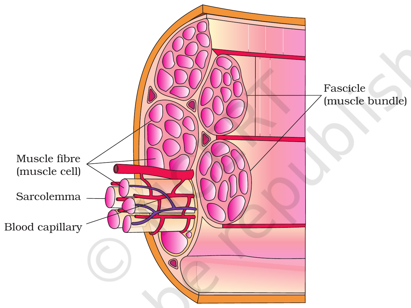
Each organised skeletal muscle in our body is made of a number of muscle bundles or fascicles held together by a common collagenous connective tissue layer called fascia. Each muscle fibre is lined by the plasma membrane called sarcolemma, enclosing the sarcoplasm. Muscle fibre is a syncitium as the sarcoplasm contains many nuclei. The sarcoplasmic reticulum of the muscle fibre is the store house of calcium ions.
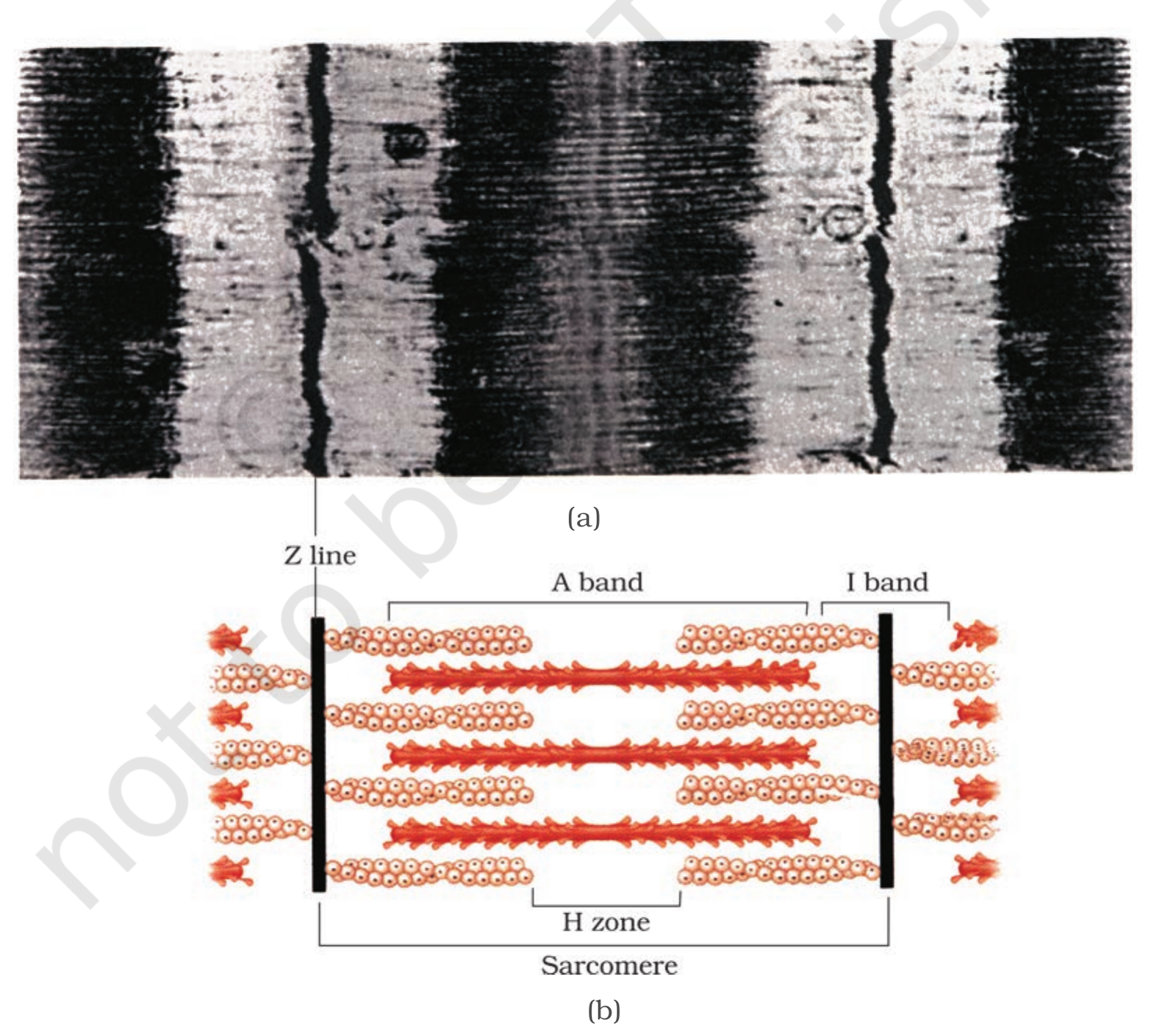
Each muscle fibre has a large number of parallelly arranged filaments in the sarcoplasm called myofibrils. Each myofibril has alternate light and dark bands. The light bands contain actin and are called I-bands or Isotropic bands, whereas the dark bands called A-bands or Anisotropic bands contain myosin.
In the centre of each I-band is an elastic fibre called the Z-line, which bisects it. The thin filaments are firmly attached to the Z-line. The thick filaments in the A-band are held together in the middle of this band by a thin fibrous membrane called the M-line. The central part of the thick filament, not overlapped by thin filaments, is called the H-zone.
The portion of the myofibril between two successive Z-lines is considered as the functional unit of contraction and is called a sarcomere.
Structure of contractile proteins
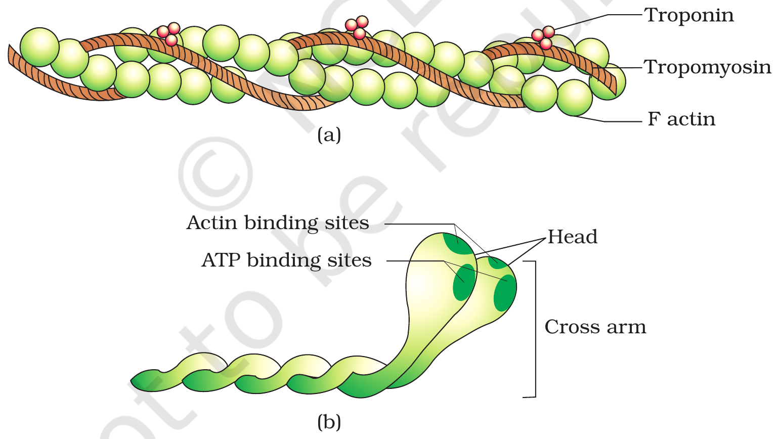
Each actin (thin) filament is made of two F-actins helically wound to each other. Each F-actin is a polymer of monomeric G-actins. Two filaments of another protein, tropomyosin, also run close to the F-actins throughout its length. A complex protein troponin is distributed at regular intervals on the tropomyosin.
Each myosin (thick) filament is also a polymerised protein. Many monomeric proteins called meromyosins constitute one thick filament. Each meromyosin has two important parts: a globular head with a short arm and a tail. The former is called the heavy meromyosin (HMM) and the latter is the light meromyosin (LMM).
The HMM component, i.e., the head and short arm, projects outwards at regular distance and angle from each other from the surface of a polymerised myosin filament and is known as the cross arm. The globular head is an active ATPase enzyme and has binding sites for ATP and active sites for actin.
Practice1 question
Try it now. A wrong answer costs nothing here, try again until you get it.
Which statement about contractile proteins is correct?
The globular head of heavy meromyosin (HMM) is an active ATPase enzyme and has binding sites for ATP and actin. Troponin is distributed at regular intervals on tropomyosin, not the other way round. Myosin is the thick filament and actin is the thin one, and F-actin (not tropomyosin) is the polymer of G-actin monomers.
Mechanism of muscle contraction
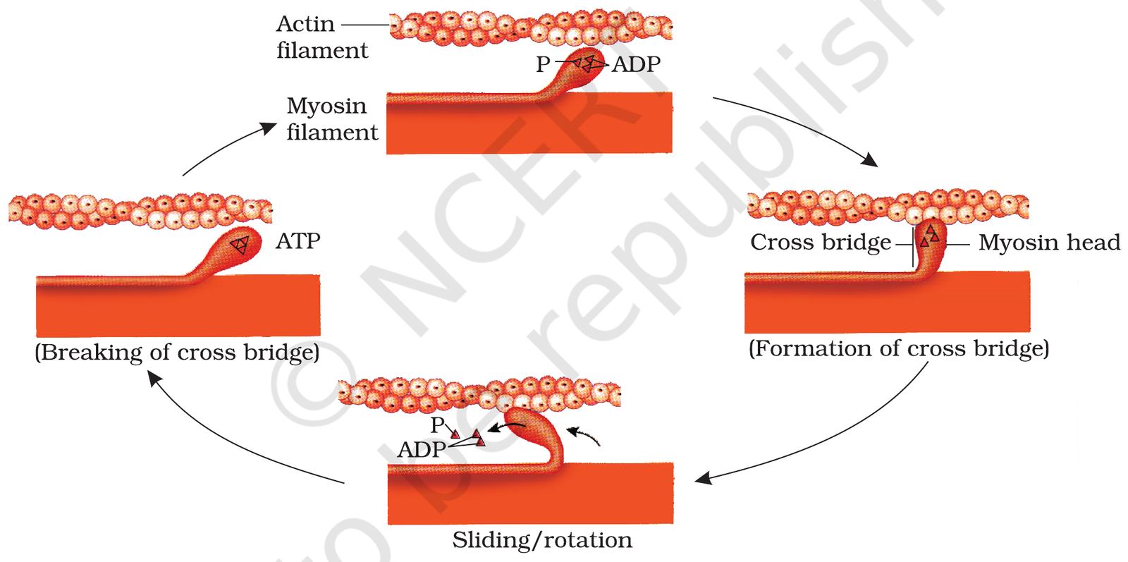
The mechanism of muscle contraction is best explained by the sliding filament theory, which states that contraction of a muscle fibre takes place by the sliding of the thin filaments over the thick filaments.
Muscle contraction is initiated by a signal sent by the central nervous system (CNS) via a motor neuron. A motor neuron along with the muscle fibres connected to it constitutes a motor unit. The junction between a motor neuron and the sarcolemma of the muscle fibre is called the neuromuscular junction or motor-end plate.
A neural signal reaching this junction releases a neurotransmitter (acetylcholine), which generates an action potential in the sarcolemma. This spreads through the muscle fibre and causes the release of calcium ions into the sarcoplasm. Increase in calcium ions initiates the sliding of thin filaments over thick filaments.
Calcium ions bind to troponin on the actin filaments. This causes tropomyosin to move away from the active sites on actin, so the active sites are exposed. The myosin head, now with energy from the hydrolysis of ATP, binds to the exposed active sites on actin to form a cross bridge. This pulls the attached actin filaments towards the centre of the A-band. The Z-lines attached to these actins are also pulled inwards, thereby causing a shortening of the sarcomere, i.e., contraction.
During contraction, the I-bands get reduced, whereas the A-bands retain their length. The H-zone disappears as the thin filaments slide inwards. ATP is used to re-energise the myosin head: the head detaches, breaks ATP again, and binds to a new active site further along the actin, so the cycle repeats as long as calcium ions are present.
When the motor neuron stops sending the signal, calcium ions are pumped back into the sarcoplasmic reticulum. This results in the masking of the active sites of actin and the myosin head detaches, so the muscle relaxes.
Red and white muscle fibres
Muscle fibres are of two types, red fibres and white fibres, based on the amount of myoglobin and mitochondria they contain.
- Red fibres: the muscle fibres are rich in myoglobin, which gives them a red colour. They contain plenty of mitochondria, which can utilise the large amount of oxygen stored in myoglobin for ATP production. They also contain more sarcoplasm. These muscles are also called aerobic muscles (Extra information: NCERT calls them aerobic muscles but does not call them slow fibres).
- White fibres: the muscle fibres contain very less quantity of myoglobin, hence they are pale or white. They contain very few mitochondria but a large amount of sarcoplasmic reticulum. They depend on anaerobic processes for energy (Extra information: NCERT does not call them fast fibres).
In coaching material, red fibres are described as slow fibres that work steadily without tiring, and white fibres as fast fibres that work powerfully in short bursts (Extra information: NCERT 17.2 only says the reaction time of the fibres can vary in different muscles).
Axial skeleton: skull and vertebral column
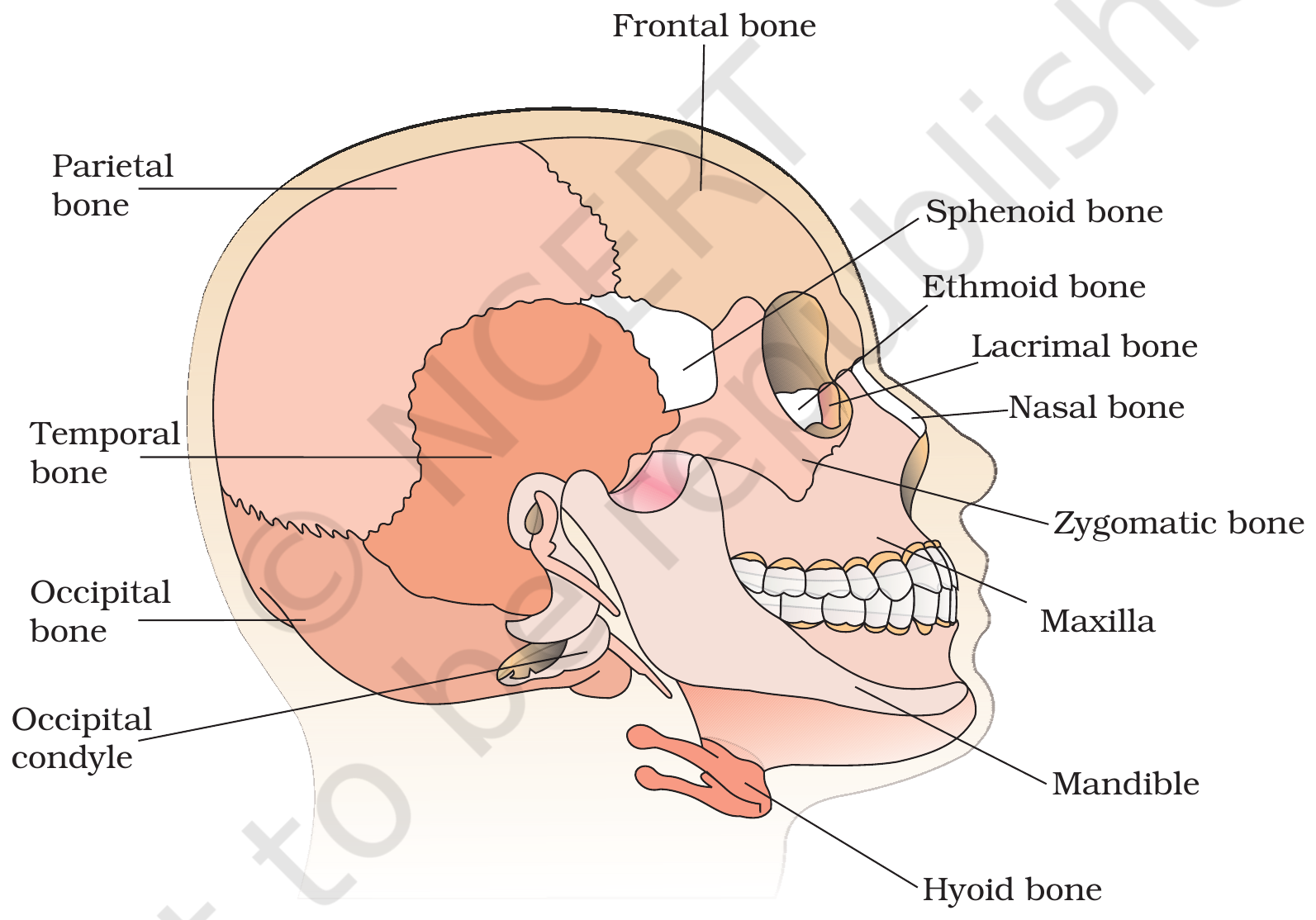
The skeletal system consists of a framework of bones and a few cartilages. This system has a significant role in movement shown by the body. Bone and cartilage are specialised connective tissues. The former has a very hard matrix due to calcium salts in it and the latter has slightly pliable matrix due to chondroitin salts.
In human beings, the skeletal system is made up of 206 bones and a few cartilages, and it is divided into two principal divisions: the axial and the appendicular skeleton. The axial skeleton comprises 80 bones distributed along the main axis of the body. The appendicular skeleton comprises 126 bones.
The skull, vertebral column, sternum and ribs constitute the axial skeleton.
- Skull: it is composed of two sets of bones, cranial and facial, that total 22 bones. The cranial bones are 8 in number. They form the hard protective outer covering, the cranium, for the brain. The facial region is made up of 14 skeletal elements which form the front part of the skull. A single U-shaped bone called hyoid is present at the base of the buccal cavity. Each middle ear contains three tiny bones: malleus, incus and stapes, collectively called ear ossicles. The skull region articulates with the superior region of the vertebral column with the help of two occipital condyles (dicondylic skull).
- Vertebral column: it is formed by 26 serially arranged units called vertebrae and is dorsally placed. It extends from the base of the skull and constitutes the main framework of the trunk. Each vertebra has a central hollow portion (the neural canal) through which the spinal cord passes. The first vertebra is the atlas and it articulates with the occipital condyles. The vertebral column is differentiated into cervical (7), thoracic (12), lumbar (5), sacral (1-fused) and coccygeal (1-fused) (Extra information: the sacral vertebra is formed by 5 fused vertebrae and the coccygeal by 4 fused vertebrae; NCERT gives only the 1-fused count.) regions, starting from the skull. The number of cervical vertebrae is seven in almost all mammals. The vertebral column protects the spinal cord, supports the head and serves as the point of attachment for the ribs and musculature of the back.
Practice1 question
Try it now. A wrong answer costs nothing here, try again until you get it.
How many bones does the axial skeleton comprise?
The axial skeleton comprises 80 bones: skull 22 (8 cranial + 14 facial), vertebral column 26, sternum 1 and ribs 24, plus the hyoid (1) and the ear ossicles (6). The appendicular skeleton has 126 bones, giving 206 bones in all.
Sternum and ribs
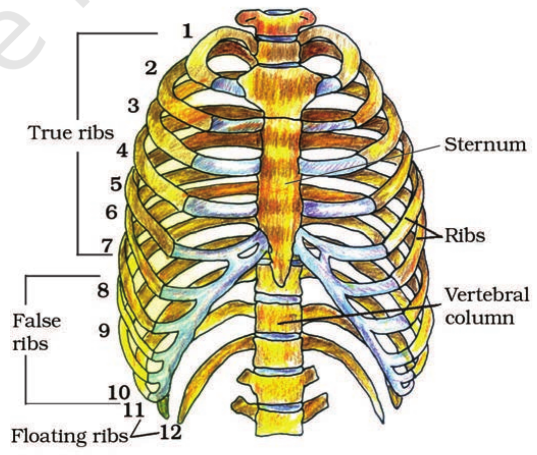
- Sternum: it is a flat bone on the ventral midline of the thorax.
- Ribs: there are 12 pairs of ribs. Each rib is a thin flat bone connected dorsally to the vertebral column and ventrally to the sternum. It has two articulation surfaces on its dorsal end and is hence called bicephalic.
- True ribs: the first seven pairs of ribs are called true ribs. Dorsally, they are attached to the thoracic vertebrae and ventrally connected to the sternum with the help of hyaline cartilage.
- False (vertebrochondral) ribs: the 8th, 9th and 10th pairs of ribs do not articulate directly with the sternum but join the 7th rib with the help of hyaline cartilage.
- Floating ribs: the last two pairs (11th and 12th) of ribs are not connected ventrally and are therefore called floating ribs.
- Rib cage: the vertebral column, sternum and ribs together form the rib cage, which encloses and protects the heart and lungs.
Appendicular skeleton: pectoral girdle and arm
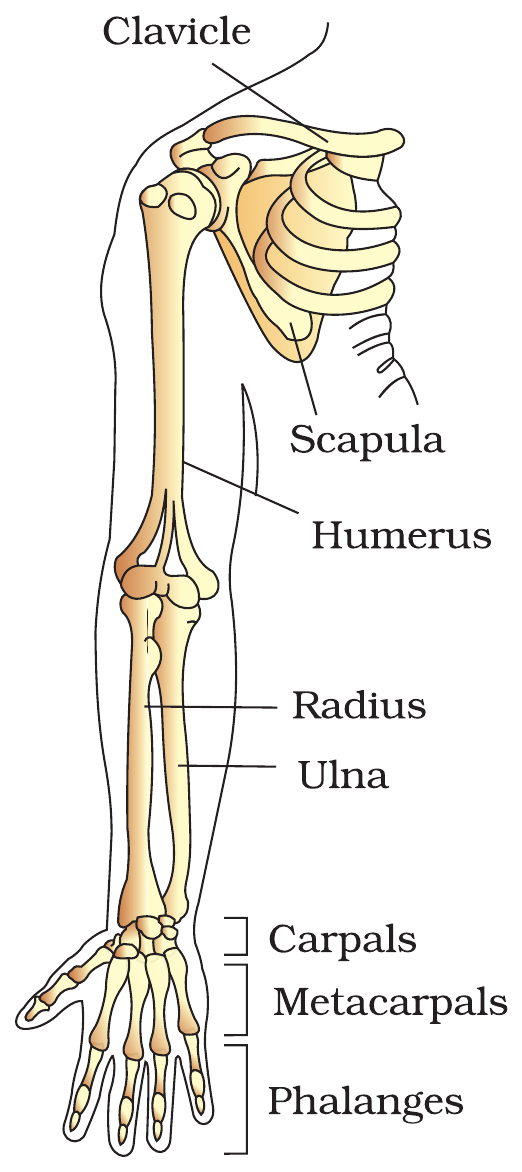
Each pectoral girdle (shoulder girdle) consists of a clavicle and a scapula. The clavicle is a long slender bone with two curvatures, commonly called the collar bone. The scapula is a large triangular flat bone situated in the dorsal part of the thorax between the second and the seventh ribs.
The dorsal, flat, triangular body of the scapula has a slightly elevated ridge called the spine, which projects as a flat, expanded process called the acromion. The clavicle articulates with this. Below the acromion is a depression called the glenoid cavity, which articulates with the head of the humerus to form the shoulder joint.
Each arm has 30 bones: humerus (1, the upper arm), radius (1) and ulna (1, the forearm), carpals (8, the wrist), metacarpals (5, the palm) and phalanges (14, the digits).
Appendicular skeleton: pelvic girdle and leg
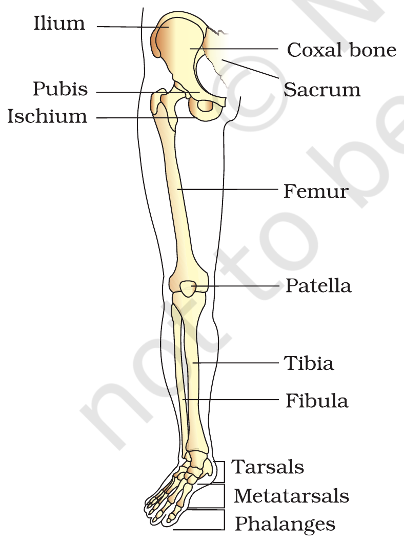
The pelvic girdle is formed by two halves, each of which is formed by the fusion of three bones: ilium, ischium and pubis. At the point of fusion of the above bones is a cavity called the acetabulum, to which the thigh bone articulates. The two halves of the pelvic girdle meet ventrally to form the pubic symphysis containing fibrous cartilage.
Each leg has 30 bones: femur (1, the thigh, the longest bone of the body) (Extra information: NCERT calls the femur the longest bone only; strongest is not in NCERT.), patella (1, a cup-shaped bone covering the knee ventrally) (Extra information: the patella is an example of a sesamoid bone, a bone formed by ossification in a tendon; NCERT does not use the term sesamoid.), tibia (1) and fibula (1) (Extra information: the tibia and fibula together are called the shank; NCERT does not use the word shank.), tarsals (7, the ankle), metatarsals (5) and phalanges (14, the digits).
Practice1 question
Try it now. A wrong answer costs nothing here, try again until you get it.
Which bone is the knee cap?
The patella is the cup-shaped bone that covers the knee ventrally; it is called the knee cap (it is a sesamoid bone, a bone formed in a tendon, which is extra information beyond NCERT). The femur is the thigh bone, the longest bone of the body; the tibia and fibula are the bones of the lower leg.
Joints
Joints are points of contact between bones, or between bones and cartilages. Joints have been classified into three major structural forms:
- Fibrous joints: they do not allow any movement. This type of joint is shown by the flat skull bones, which fuse end-to-end with the help of dense fibrous connective tissues in the form of sutures, to form the cranium.
- Cartilaginous joints: the bones involved are joined together with the help of cartilages. The joint between the adjacent vertebrae in the vertebral column is of this pattern and it permits limited movements.
- Synovial joints: these are characterised by the presence of a fluid-filled synovial cavity between the articulating surfaces of the two bones. Such an arrangement allows considerable movement, and these joints help in locomotion and many other movements. Ball and socket joints (between the humerus and pectoral girdle), hinge joints (knee joint), pivot joints (between the atlas and axis), gliding joints (between the carpals) and saddle joints (between the carpal and metacarpal of the thumb) are some examples of synovial joints. (Extra information: the femur and acetabulum as a ball and socket joint and the elbow as a hinge joint are not in NCERT 17.4; the femur-acetabulum example appears in the chapter Exercises.)
Practice1 question
Try it now. A wrong answer costs nothing here, try again until you get it.
Which is an example of a pivot joint?
The pivot joint permits rotation and is found between the atlas and the axis vertebrae. The knee is a hinge joint, the shoulder is a ball and socket joint, and the joints between carpals are gliding joints. (Extra information: the elbow is also a hinge joint; NCERT 17.4 names only the knee.)
Disorders of muscular and skeletal system
- Myasthenia gravis: an autoimmune disorder affecting the neuromuscular junction, leading to fatigue, weakening and paralysis of skeletal muscles.
- Muscular dystrophy: progressive degeneration of skeletal muscle, mostly due to genetic disorder.
- Tetany: rapid spasms in muscle due to low $Ca^+$+ in body fluid.
- Arthritis: inflammation of joints.
- Osteoporosis: an age-related disorder characterised by decreased bone mass and increased chances of fractures. Decreased levels of estrogen is a common cause.
- Gout: inflammation of joints due to accumulation of uric acid crystals.
Table 1 - Three types of muscle
| Feature | Skeletal muscle | Visceral (smooth) muscle | Cardiac muscle |
|---|---|---|---|
| Appearance | Striated | Non-striated | Striated |
| Control | Voluntary | Involuntary | Involuntary |
| Location | Attached to skeletal bones | Walls of hollow visceral organs (alimentary canal, reproductive tract) | Heart only |
| Fibre | Syncitium, many nuclei | Smooth in appearance; fusiform (spindle-shaped) and uninucleate (extra information) | Branched cells, interconnected |
| Example | Skeletal muscle of the limbs | Alimentary canal, reproductive tract | Heart |
Source: NCERT XI, Ch 17, 17.2.
Table 2 - Axial vs appendicular skeleton
| Division | Parts | Bones |
|---|---|---|
| Axial (80) | Skull | 22 cranial + facial; hyoid 1; ear ossicles 6 |
| Axial (80) | Vertebral column | 26 (cervical 7, thoracic 12, lumbar 5, sacral 1, coccygeal 1) |
| Axial (80) | Sternum | 1 |
| Axial (80) | Ribs | 24 (12 pairs) |
| Appendicular (126) | Pectoral girdles | 4 (clavicle 2, scapula 2) |
| Appendicular (126) | Upper limbs | 60 (30 in each limb) |
| Appendicular (126) | Pelvic girdle | 2 coxal bones |
| Appendicular (126) | Lower limbs | 60 (30 in each limb) |
Source: NCERT XI, Ch 17, 17.3.
Table 3 - Joints at a glance
| Joint type | Movement allowed | Example |
|---|---|---|
| Fibrous | No movement | Flat skull bones (sutures) |
| Cartilaginous | Limited movement | Between adjacent vertebrae |
| Ball and socket (synovial) | Free movement in many directions | Humerus and pectoral girdle (extra information: femur and acetabulum) |
| Hinge (synovial) | Movement in one plane | Knee joint (extra information: elbow joint) |
| Pivot (synovial) | Rotation | Between atlas and axis |
| Gliding (synovial) | Sliding movement | Between carpals |
| Saddle (synovial) | Movement in two planes | Between carpal and metacarpal of thumb |
Source: NCERT XI, Ch 17, 17.4.
Ladder - The confusions that cost marks
| Trap | Wrong | Right |
|---|---|---|
| A band | The A band shortens during contraction | The A band stays the same length; the I band shortens and the H zone disappears |
| Troponin and tropomyosin | Tropomyosin is distributed on troponin | Troponin is distributed at regular intervals on tropomyosin |
| Ribs | Ribs 8 to 10 are floating ribs | Ribs 8 to 10 are false (vertebrochondral) ribs; ribs 11 and 12 are floating |
| Red and white fibres | Red fibres contract fast | Red fibres are slow and aerobic (myoglobin rich); white fibres are fast and anaerobic |
| Joints between vertebrae | The joint between adjacent vertebrae is fibrous | It is cartilaginous and permits limited movement |
| Tetany and gout | Tetany is caused by uric acid crystals | Tetany is rapid spasm from low $Ca^+$+ in body fluid; gout is uric acid crystal accumulation in joints |
| Hinge and pivot | The shoulder is a hinge joint | The shoulder is a ball and socket joint; the hinge joint is the knee joint |
| Skull bones | The skull has 29 bones | The skull has 22 bones (8 cranial + 14 facial); hyoid 1 and ear ossicles 6 are separate |
Source: NCERT XI, Ch 17; NEET 2019, 2021, 2023, 2024.
Trap Boxes
The A band shortens during contraction?
Wrong idea: During muscle contraction the A band shortens and the I band stays the same.
The A band retains its length during contraction. The I bands get reduced and the H zone disappears as the thin filaments slide towards the centre of the A band. NEET 2021.
NCERT XI, Ch 17, 17.2.2
Tropomyosin is distributed on troponin?
Wrong idea: Tropomyosin is distributed at regular intervals on troponin.
Troponin is distributed at regular intervals on tropomyosin. Tropomyosin runs close to the F-actin filaments. NEET 2023 asked exactly this reversal.
NCERT XI, Ch 17, 17.2.1
Ribs 8 to 10 are floating ribs?
Wrong idea: The last five pairs of ribs, 8 to 12, are all floating ribs.
Ribs 1 to 7 are true ribs, ribs 8 to 10 are false (vertebrochondral) ribs joined to the 7th rib, and ribs 11 and 12 are floating ribs, not connected ventrally.
NCERT XI, Ch 17, 17.3
Red fibres contract fast?
Wrong idea: Red muscle fibres contract fast because they have plenty of mitochondria.
Red fibres are rich in myoglobin and mitochondria, use aerobic respiration and contract slowly. White fibres have very little myoglobin and few mitochondria, depend on anaerobic processes and contract fast.
NCERT XI, Ch 17, 17.2
Tetany is caused by uric acid?
Wrong idea: Tetany is the inflammation of joints caused by uric acid crystals.
Tetany is rapid spasm in muscle due to low $Ca^+$+ in the body fluid. Gout is inflammation of joints due to accumulation of uric acid crystals.
NCERT XI, Ch 17, 17.5
Chapter test
5 NEET PYQs. +4 for a correct answer, -1 for a wrong answer, exactly like NEET. Finish the test to earn a medal.
Q1: NEET (UG) 2024, Code T1, Q166
Match List I with List II:
| List I | List II |
|---|---|
| A. Fibrous joints | I. Adjacent vertebrae, limited movement |
| B. Cartilaginous joints | II. Humerus and Pectoral girdle, rotational movement |
| C. Hinge joints | III. Skull, don't allow any movement |
| D. Ball and socket joints | IV. Knee, help in locomotion |
Fibrous joints do not allow any movement and are the skull sutures (A-III); cartilaginous joints are between adjacent vertebrae and permit limited movement (B-I); hinge joints are the knee, which helps in locomotion (C-IV); ball and socket joints are between the humerus and the pectoral girdle and allow rotational movement (D-II).
Q2: NEET (UG) 2023, Code E1, Q199
Which of the following statements are correct regarding skeletal muscle?
A. Muscle bundles are held together by collagenous connective tissue layer called fascicle.
B. Sarcoplasmic reticulum of muscle fibre is a store house of calcium ions.
C. Striated appearance of skeletal muscle fibre is due to distribution pattern of actin and myosin proteins.
D. M line is considered as functional unit of contraction called sarcomere.
Choose the most appropriate answer from the options given below:
B and C are correct: the sarcoplasmic reticulum of a muscle fibre stores calcium ions, and the striated appearance of skeletal muscle comes from the distribution pattern of actin and myosin. A is wrong because the fascicle is the muscle bundle itself, and D is wrong because the sarcomere, not the M line, is the functional unit of contraction.
Q3: NEET (UG) 2022, Code R1, Q167
Given below are two statements: one is labelled as Assertion (A) and the other is labelled as Reason (R).
Assertion (A): Osteoporosis is characterised by decreased bone mass and increased chance of fractures.
Reason (R): Common cause of osteoporosis is increased levels of estrogen.
In the light of the above statements, choose the most appropriate answer from the options given below:
Osteoporosis is an age-related disorder characterised by decreased bone mass and increased chances of fractures, so the Assertion is correct. Its common cause is decreased, not increased, levels of estrogen, so the Reason is false.
Q4: NEET (UG) 2021, Code M1, Q187
During muscular contraction which of the following events occur?
(a) 'H' zone disappears
(b) 'A' band widens
(c) 'I' band reduces in width
(d) Myosine hydrolyzes ATP, releasing the ADP and Pi
(e) Z-lines attached to actins are pulled inwards
Choose the correct answer from the options given below:
During contraction the H zone disappears, the I band reduces in width, myosin hydrolyses ATP releasing ADP and Pi, and the Z lines attached to actins are pulled inwards. The A band does not widen; it retains its length.
Q5: NEET (UG) 2019, Code S2, Q52
Select the correct option.
The first seven pairs of ribs are vertebrosternal (true ribs), attached to the sternum with hyaline cartilage; the 8th, 9th and 10th pairs are vertebrochondral (false ribs), joined to the 7th rib; the 11th and 12th pairs are vertebral (floating) ribs, not connected ventrally.
Score: 0 · Answered 0 of 5
Flashcards
Tap a card to flip it over and see the answer. Tap it again to flip it back.
How many bones are in the axial and appendicular skeletons?
Tap to flip80 bones in the axial skeleton and 126 in the appendicular skeleton: 206 bones in total in an adult.
Tap to flip backPut the steps of muscle contraction in order.
Tap to flipSignal from the CNS reaches the muscle fibre through a motor neuron, acetylcholine is released at the neuromuscular junction, an action potential spreads in the sarcolemma, calcium ions are released from the sarcoplasmic reticulum, calcium binds to troponin, the myosin head binds actin and pulls it towards the centre o
Tap to flip backWhich is the thin filament and which is the thick filament?
Tap to flipActin is the thin filament; myosin is the thick filament.
Tap to flip backWhich muscle is striated but involuntary?
Tap to flipCardiac muscle. It is striated like skeletal muscle but involuntary, found only in the heart.
Tap to flip backHow many vertebrae are in each region of the vertebral column?
Tap to flip7 cervical, 12 thoracic, 5 lumbar, 1 sacral (1-fused) and 1 coccygeal (1-fused): 26 in all. (Extra information: the sacral vertebra is formed by 5 fused vertebrae and the coccygeal by 4 fused vertebrae.)
Tap to flip backWhich ribs are true, false and floating?
Tap to flipRibs 1 to 7 are true ribs (attached to the sternum), ribs 8 to 10 are false or vertebrochondral ribs (joined to the 7th rib), ribs 11 and 12 are floating ribs (not connected ventrally).
Tap to flip backHow many bones make up the skull, and how are they divided?
Tap to flipThe skull has 22 bones: 8 cranial and 14 facial. The hyoid (1) and the ear ossicles (6) are separate.
Tap to flip backWhat is a sarcomere?
Tap to flipThe part of a myofibril between two successive Z lines. It is the functional unit of contraction.
Tap to flip backWhich joint is between the atlas and the axis?
Tap to flipThe pivot joint between the atlas and the axis vertebrae.
Tap to flip backWhat is osteoporosis, and what is a common cause?
Tap to flipDecreased bone mass and increased chances of fractures. Decreased levels of estrogen is a common cause in aged women.
Tap to flip back