 NEET Notes
Home > Biology > Botany > Cell Cycle and Cell Division
Cell Cycle and Cell Division
On this page · 18 sections
- 01Syllabus map
- 02Cell cycle
- 03Interphase: G1, S and G2
- 04Quiescent stage and mitosis in haploid cells
- 05M phase: prophase
- 06Metaphase, anaphase and telophase
- 07Cytokinesis
- 08Significance of mitosis
- 09Meiosis: key features
- 10Prophase I: five substages
- 11Metaphase I to telophase I and interkinesis
- 12Meiosis II
- 13Significance of meiosis
- 14Table 1 - Interphase phases compared
- 15Table 2 - Mitosis: key events of each stage
- 16Table 3 - Prophase I substages
- 17Table 4 - Mitosis vs meiosis
- 18Table 5 - Meiosis I vs meiosis II
The cell cycle, mitosis and meiosis: interphase phases, the four stages of mitosis, the five substages of prophase I, and why meiosis halves the chromosome number.
Syllabus map
- NMC Unit: BIO-U03, Cell Structure and Function. NCERT Chapter 10, Cell Cycle and Cell Division.
- Subtopics in this chapter (NCERT XI, Ch 10): Cell cycle and its phases. M phase. Significance of mitosis. Meiosis. Significance of meiosis.
- Subtopic checklist: (1) phases of the cell cycle, G1, S, G2 and M; (2) DNA content 2C to 4C in S phase; (3) the four stages of mitosis and cytokinesis; (4) the five substages of prophase I; (5) meiosis I and meiosis II; (6) why mitosis is equational and meiosis is reductional.
- Excluded topics: detail of cell structure (Chapter 8, Cell: The Unit of Life) and biomolecules (Chapter 9, Biomolecules). Those are covered in their own chapters; only the facts this chapter needs are repeated here.
- Estimated revision time: 14 minutes. Jump links: Syllabus Map, Cell Cycle, Interphase, M Phase, Cytokinesis, Meiosis, Trap Boxes, PYQs, Chapter Test.
Cell cycle
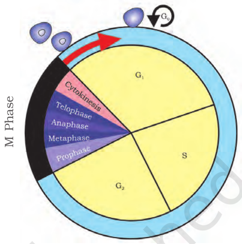
All organisms, even the largest, start their life from a single cell. Growth and reproduction are characteristics of cells, indeed of all living organisms. All cells reproduce by dividing into two, with each parental cell giving rise to two daughter cells each time they divide.
Cell division is a very important process in all living organisms. During the division of a cell, DNA replication and cell growth also take place. All these processes, i.e., cell division, DNA replication, and cell growth, have to take place in a coordinated way to ensure correct division and formation of progeny cells containing intact genomes.
The sequence of events by which a cell duplicates its genome, synthesises the other constituents of the cell and eventually divides into two daughter cells is termed cell cycle. Although cell growth (in terms of cytoplasmic increase) is a continuous process, DNA synthesis occurs only during one specific stage in the cell cycle. The replicated chromosomes (DNA) are then distributed to daughter nuclei by a complex series of events during cell division. These events are themselves under genetic control.
The cell cycle is divided into two basic phases: Interphase and M Phase (Mitosis phase). The M Phase represents the phase when the actual cell division or mitosis occurs, and the interphase represents the phase between two successive M phases.
A typical eukaryotic cell cycle is illustrated by human cells in culture. These cells divide once in approximately every 24 hours. However, this duration of cell cycle can vary from organism to organism and also from cell type to cell type. Yeast, for example, can progress through the cell cycle in only about 90 minutes. In the 24 hour average duration of cell cycle of a human cell, cell division proper lasts for only about an hour. The interphase lasts more than 95% of the duration of cell cycle.
Interphase: G1, S and G2
The M Phase starts with the nuclear division, corresponding to the separation of daughter chromosomes (karyokinesis) and usually ends with division of cytoplasm (cytokinesis). The interphase, though called the resting phase, is the time during which the cell is preparing for division by undergoing both cell growth and DNA replication in an orderly manner.
The interphase is divided into three further phases:
- G1 phase (Gap 1): G1 phase corresponds to the interval between mitosis and initiation of DNA replication. During G1 phase the cell is metabolically active and continuously grows but does not replicate its DNA.
- S phase (Synthesis): S or synthesis phase marks the period during which DNA synthesis or replication takes place. During this time the amount of DNA per cell doubles. If the initial amount of DNA is denoted as 2C then it increases to 4C. However, there is no increase in the chromosome number; if the cell had diploid or 2n number of chromosomes at G1, even after S phase the number of chromosomes remains the same, i.e., 2n.
- G2 phase (Gap 2): During the G2 phase, proteins are synthesised in preparation for mitosis while cell growth continues.
In animal cells, during the S phase, DNA replication begins in the nucleus, and the centriole duplicates in the cytoplasm.
Worked example from NCERT: mitosis in onion root tip cells. It has 16 chromosomes in each cell. The cell has 16 chromosomes at G1 phase, 16 chromosomes after S phase, and 16 chromosomes after M phase. If the DNA content after M phase is 2C, the DNA content is 2C at G1, 4C after S phase, and 4C at G2.
Practice1 question
Try it now. A wrong answer costs nothing here, try again until you get it.
A diploid cell has 2C DNA at G1. What are its DNA content and chromosome number at G2?
DNA doubles in S phase: 2C becomes 4C. The chromosome number does not change in S phase, so the cell stays 2n.
Quiescent stage and mitosis in haploid cells
Some cells in the adult animals do not appear to exhibit division (e.g., heart cells) and many other cells divide only occasionally, as needed to replace cells that have been lost because of injury or cell death. These cells that do not divide further exit G1 phase to enter an inactive stage called quiescent stage (G0) of the cell cycle. Cells in this stage remain metabolically active but no longer proliferate unless called on to do so depending on the requirement of the organism.
In animals, mitotic cell division is only seen in the diploid somatic cells. However, there are few exceptions to this where haploid cells divide by mitosis, for example, male honey bees. Against this, the plants can show mitotic divisions in both haploid and diploid cells.
M phase: prophase

M Phase is the most dramatic period of the cell cycle, involving a major reorganisation of virtually all components of the cell. Since the number of chromosomes in the parent and progeny cells is the same, it is also called as equational division.
Though for convenience mitosis has been divided into four stages of nuclear division (karyokinesis), it is very essential to understand that cell division is a progressive process and very clear-cut lines cannot be drawn between various stages. Karyokinesis involves four stages: Prophase, Metaphase, Anaphase and Telophase.
Prophase, the first stage of karyokinesis of mitosis, follows the S and G2 phases of interphase. In the S and G2 phases the new DNA molecules formed are not distinct but intertwined. Prophase is marked by the initiation of condensation of chromosomal material. The chromosomal material becomes untangled during the process of chromatin condensation. The centrosome, which had undergone duplication during S phase of interphase, now begins to move towards opposite poles of the cell.
The completion of prophase can thus be marked by the following characteristic events:
- Chromosomal material condenses to form compact mitotic chromosomes. Chromosomes are seen to be composed of two chromatids attached together at the centromere.
- Centrosome which had undergone duplication during interphase, begins to move towards opposite poles of the cell. Each centrosome radiates out microtubules called asters. The two asters together with spindle fibres form the mitotic apparatus.
- Cells at the end of prophase, when viewed under the microscope, do not show golgi complexes, endoplasmic reticulum, nucleolus and the nuclear envelope.
Practice1 question
Try it now. A wrong answer costs nothing here, try again until you get it.
Which of these marks the end of prophase in mitosis?
By the end of prophase the golgi complexes, endoplasmic reticulum, nucleolus and nuclear envelope are no longer visible. The chromosomes have condensed and the centrosomes have moved to opposite poles.
Metaphase, anaphase and telophase
Metaphase: the complete disintegration of the nuclear envelope marks the start of the second phase of mitosis, hence the chromosomes are spread through the cytoplasm of the cell. By this stage, condensation of chromosomes is completed and they can be observed clearly under the microscope. This then, is the stage at which morphology of chromosomes is most easily studied. At this stage, metaphase chromosome is made up of two sister chromatids, which are held together by the centromere. Small disc-shaped structures at the surface of the centromeres are called kinetochores. These structures serve as the sites of attachment of spindle fibres to the chromosomes that are moved into position at the centre of the cell. Hence, the metaphase is characterised by all the chromosomes coming to lie at the equator with one chromatid of each chromosome connected by its kinetochore to spindle fibres from one pole and its sister chromatid connected by its kinetochore to spindle fibres from the opposite pole. The plane of alignment of the chromosomes at metaphase is referred to as the metaphase plate. The key features of metaphase are: spindle fibres attach to kinetochores of chromosomes, and chromosomes are moved to spindle equator and get aligned along metaphase plate through spindle fibres to both poles.
Anaphase: at the onset of anaphase, each chromosome arranged at the metaphase plate is split simultaneously and the two daughter chromatids, now referred to as daughter chromosomes of the future daughter nuclei, begin their migration towards the two opposite poles. As each chromosome moves away from the equatorial plate, the centromere of each chromosome remains directed towards the pole and hence at the leading edge, with the arms of the chromosome trailing behind. Thus, anaphase stage is characterised by the following key events: centromeres split and chromatids separate, and chromatids move to opposite poles.
Telophase: at the beginning of the final stage of karyokinesis, i.e., telophase, the chromosomes that have reached their respective poles decondense and lose their individuality. The individual chromosomes can no longer be seen and each set of chromatin material tends to collect at each of the two poles. This is the stage which shows the following key events: chromosomes cluster at opposite spindle poles and their identity is lost as discrete elements; nuclear envelope develops around the chromosome clusters at each pole forming two daughter nuclei; nucleolus, golgi complex and ER reform.
Practice1 question
Try it now. A wrong answer costs nothing here, try again until you get it.
At which stage of mitosis do all chromosomes lie at the equator?
Metaphase is characterised by all chromosomes coming to lie at the equator, on the metaphase plate. Chromosome morphology is most easily studied at this stage.
Cytokinesis
Mitosis accomplishes not only the segregation of duplicated chromosomes into daughter nuclei (karyokinesis), but the cell itself is divided into two daughter cells by the separation of cytoplasm called cytokinesis, at the end of which cell division gets completed.
In an animal cell, cytokinesis is achieved by the appearance of a furrow in the plasma membrane. The furrow gradually deepens and ultimately joins in the centre dividing the cell cytoplasm into two.
Plant cells however, are enclosed by a relatively inextensible cell wall, therefore they undergo cytokinesis by a different mechanism. In plant cells, wall formation starts in the centre of the cell and grows outward to meet the existing lateral walls. The formation of the new cell wall begins with the formation of a simple precursor, called the cell-plate that represents the middle lamella between the walls of two adjacent cells.
At the time of cytoplasmic division, organelles like mitochondria and plastids get distributed between the two daughter cells. In some organisms karyokinesis is not followed by cytokinesis as a result of which multinucleate condition arises leading to the formation of syncytium (e.g., liquid endosperm in coconut).
Significance of mitosis
Mitosis or the equational division is usually restricted to the diploid cells only. However, in some lower plants and in some social insects haploid cells also divide by mitosis.
Mitosis usually results in the production of diploid daughter cells with identical genetic complement. The growth of multicellular organisms is due to mitosis. Cell growth results in disturbing the ratio between the nucleus and the cytoplasm. It therefore becomes essential for the cell to divide to restore the nucleo-cytoplasmic ratio.
A very significant contribution of mitosis is cell repair. The cells of the upper layer of the epidermis, cells of the lining of the gut, and blood cells are being constantly replaced. Mitotic divisions in the meristematic tissues - the apical and the lateral cambium, result in a continuous growth of plants throughout their life.
Meiosis: key features
The production of offspring by sexual reproduction includes the fusion of two gametes, each with a complete haploid set of chromosomes. Gametes are formed from specialised diploid cells. This specialised kind of cell division that reduces the chromosome number by half results in the production of haploid daughter cells. This kind of division is called meiosis. Meiosis ensures the production of haploid phase in the life cycle of sexually reproducing organisms whereas fertilisation restores the diploid phase. We come across meiosis during gametogenesis in plants and animals. This leads to the formation of haploid gametes.
The key features of meiosis are as follows:
- Meiosis involves two sequential cycles of nuclear and cell division called meiosis I and meiosis II but only a single cycle of DNA replication.
- Meiosis I is initiated after the parental chromosomes have replicated to produce identical sister chromatids at the S phase.
- Meiosis involves pairing of homologous chromosomes and recombination between non-sister chromatids of homologous chromosomes.
- Four haploid cells are formed at the end of meiosis II.
Meiotic events can be grouped under the following phases: Meiosis I - Prophase I, Metaphase I, Anaphase I, Telophase I; Meiosis II - Prophase II, Metaphase II, Anaphase II, Telophase II.
Prophase I: five substages
Prophase of the first meiotic division is typically longer and more complex when compared to prophase of mitosis. It has been further subdivided into the following five phases based on chromosomal behaviour, i.e., Leptotene, Zygotene, Pachytene, Diplotene and Diakinesis.
- Leptotene: during leptotene stage the chromosomes become gradually visible under the light microscope. The compaction of chromosomes continues throughout leptotene.
- Zygotene: during this stage chromosomes start pairing together and this process of association is called synapsis. Such paired chromosomes are called homologous chromosomes. Electron micrographs of this stage indicate that chromosome synapsis is accompanied by the formation of complex structure called synaptonemal complex. The complex formed by a pair of synapsed homologous chromosomes is called a bivalent or a tetrad. However, these are more clearly visible at the next stage. The first two stages of prophase I are relatively short-lived compared to the next stage that is pachytene.
- Pachytene: during this stage, the four chromatids of each bivalent chromosomes becomes distinct and clearly appears as tetrads. This stage is characterised by the appearance of recombination nodules, the sites at which crossing over occurs between non-sister chromatids of the homologous chromosomes. Crossing over is the exchange of genetic material between two homologous chromosomes. Crossing over is also an enzyme-mediated process and the enzyme involved is called recombinase. Crossing over leads to recombination of genetic material on the two chromosomes. Recombination between homologous chromosomes is completed by the end of pachytene, leaving the chromosomes linked at the sites of crossing over.
- Diplotene: the beginning of diplotene is recognised by the dissolution of the synaptonemal complex and the tendency of the recombined homologous chromosomes of the bivalents to separate from each other except at the sites of crossovers. These X-shaped structures, are called chiasmata. In oocytes of some vertebrates, diplotene can last for months or years.
- Diakinesis: the final stage of meiotic prophase I is diakinesis. This is marked by terminalisation of chiasmata. During this phase the chromosomes are fully condensed and the meiotic spindle is assembled to prepare the homologous chromosomes for separation. By the end of diakinesis, the nucleolus disappears and the nuclear envelope also breaks down. Diakinesis represents transition to metaphase.
Practice1 question
Try it now. A wrong answer costs nothing here, try again until you get it.
In which substage of prophase I does synapsis (pairing of homologous chromosomes) take place?
Synapsis is the pairing of homologous chromosomes, and it happens in zygotene, the second substage of prophase I. The synaptonemal complex forms at the same time.
Metaphase I to telophase I and interkinesis
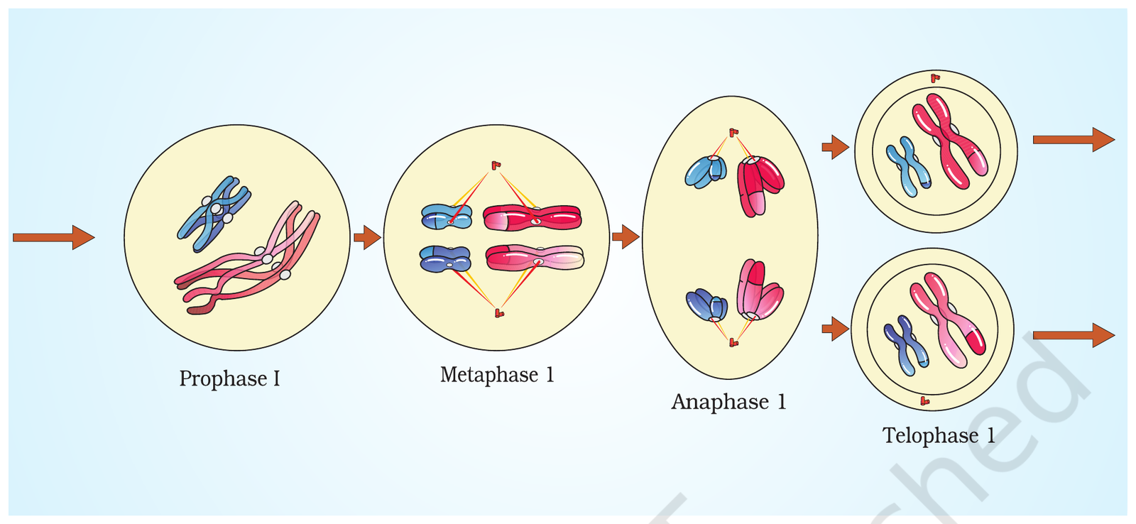
Metaphase I: the bivalent chromosomes align on the equatorial plate. The microtubules from the opposite poles of the spindle attach to the kinetochore of homologous chromosomes.
Anaphase I: the homologous chromosomes separate, while sister chromatids remain associated at their centromeres.
Telophase I: the nuclear membrane and nucleolus reappear, cytokinesis follows and this is called as dyad of cells. Although in many cases the chromosomes do undergo some dispersion, they do not reach the extremely extended state of the interphase nucleus.
The stage between the two meiotic divisions is called interkinesis and is generally short lived. There is no replication of DNA during interkinesis. Interkinesis is followed by prophase II, a much simpler prophase than prophase I.
Meiosis II
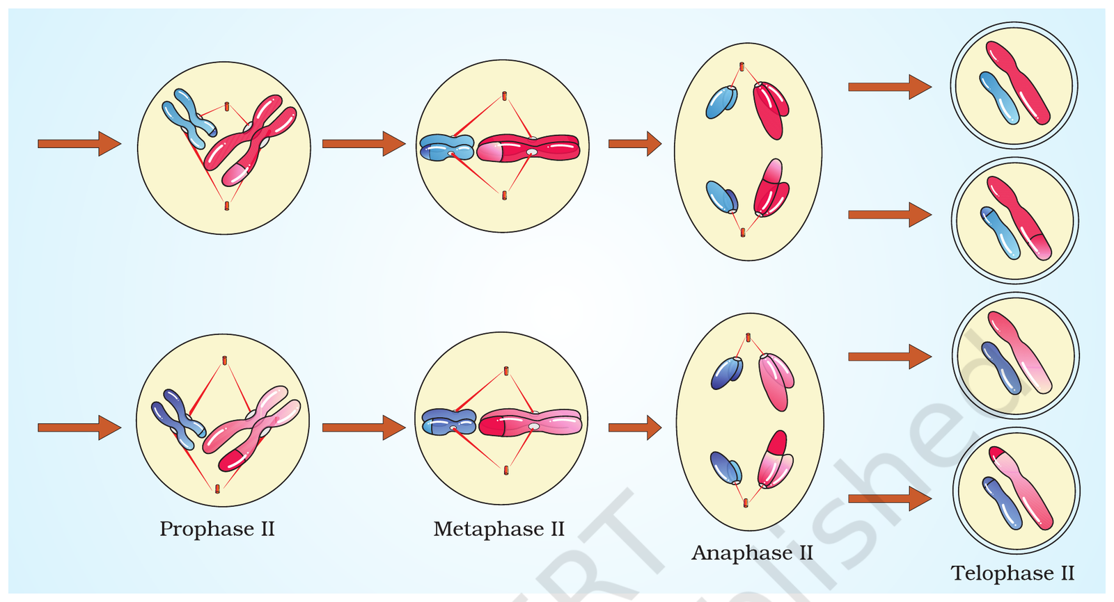
Meiosis II is initiated immediately after cytokinesis, usually before the chromosomes have fully elongated. In contrast to meiosis I, meiosis II resembles a normal mitosis.
- Prophase II: the nuclear membrane disappears by the end of prophase II. The chromosomes again become compact.
- Metaphase II: at this stage the chromosomes align at the equator and the microtubules from opposite poles of the spindle get attached to the kinetochores of sister chromatids.
- Anaphase II: it begins with the simultaneous splitting of the centromere of each chromosome (which was holding the sister chromatids together), allowing them to move toward opposite poles of the cell by shortening of microtubules attached to kinetochores.
- Telophase II: meiosis ends with telophase II, in which the two groups of chromosomes once again get enclosed by a nuclear envelope; cytokinesis follows resulting in the formation of tetrad of cells i.e., four haploid daughter cells.
Practice1 question
Try it now. A wrong answer costs nothing here, try again until you get it.
In which stage does the centromere of each chromosome split during meiosis?
Meiosis II is like mitosis: the centromere splits in anaphase II and sister chromatids separate. In anaphase I it is the homologous chromosomes that separate, while sister chromatids stay together.
Significance of meiosis
Meiosis is the mechanism by which conservation of specific chromosome number of each species is achieved across generations in sexually reproducing organisms, even though the process, per se, paradoxically, results in reduction of chromosome number by half.
It also increases the genetic variability in the population of organisms from one generation to the next. Variations are very important for the process of evolution.
The genetic variability comes from crossing over between non-sister chromatids of homologous chromosomes during pachytene. Meiosis maintains the chromosome number from generation to generation: it reduces the chromosome number to half so that the process of fertilisation restores the original number in the zygote. Extra information (beyond NCERT XI Ch 10): the random distribution of homologous chromosomes between the daughter cells at anaphase I also adds variability.
Table 1 - Interphase phases compared
| Phase | What happens | DNA content | Chromosome number |
|---|---|---|---|
| G1 (Gap 1) | Cell grows, metabolically active, no DNA replication | 2C | 2n |
| S (Synthesis) | DNA replication; centriole duplicates in animal cells | 2C to 4C | 2n (no change) |
| G2 (Gap 2) | Proteins synthesised for mitosis; cell growth continues | 4C | 2n |
| G0 (Quiescent) | Cells exit G1 and do not divide further unless called on; metabolically active | 2C | 2n |
Source: NCERT XI, Ch 10, 10.1.1. Remember: DNA doubles in S phase, chromosome number does not.
Table 2 - Mitosis: key events of each stage
| Stage | Key events |
|---|---|
| Prophase | Chromosomes condense; two chromatids joined at centromere; centrosomes move to opposite poles; asters and spindle form; golgi, ER, nucleolus and nuclear envelope disappear |
| Metaphase | Nuclear envelope disintegrates; chromosomes align at the equator on the metaphase plate; spindle fibres attach to kinetochores; chromosome morphology best studied here |
| Anaphase | Centromeres split; sister chromatids separate and move to opposite poles; centromere leads, arms trail |
| Telophase | Chromosomes decondense and lose individuality; nuclear envelope reforms; nucleolus, golgi complex and ER reform; two daughter nuclei form |
| Cytokinesis | Cytoplasm divides: furrow in animal cells, cell plate in plant cells |
Source: NCERT XI, Ch 10, 10.2.
Table 3 - Prophase I substages
| Substage | Key events |
|---|---|
| Leptotene | Chromosomes become gradually visible; compaction continues |
| Zygotene | Synapsis: homologous chromosomes pair; synaptonemal complex forms; bivalent (tetrad) forms |
| Pachytene | Tetrads clearly visible; recombination nodules appear; crossing over between non-sister chromatids (enzyme recombinase); recombination completed |
| Diplotene | Synaptonemal complex dissolves; homologues separate except at chiasmata (X-shaped); can last for months or years in oocytes of some vertebrates |
| Diakinesis | Terminalisation of chiasmata; chromosomes fully condensed; spindle assembled; nucleolus disappears; nuclear envelope breaks down |
Source: NCERT XI, Ch 10, 10.4.1.
Table 4 - Mitosis vs meiosis
| Feature | Mitosis | Meiosis |
|---|---|---|
| Divisions | One division | Two divisions: meiosis I and meiosis II |
| DNA replication | Once, before division | Once only, before meiosis I |
| Daughter cells | Two genetically identical cells (usually diploid; haploid in male honey bees and haploid plant cells) | Four haploid cells |
| Chromosome number | Same as parent (equational division) | Halved (reduction division) |
| Homologous pairing | No pairing, no crossing over | Pairing (synapsis) and crossing over in prophase I |
| Prophase | Short, no substages | Prophase I is long, with five substages |
| Anaphase | Centromeres split, chromatids separate | Anaphase I: homologues separate; anaphase II: centromeres split |
| Purpose | Growth, repair, restoring nucleo-cytoplasmic ratio | Gamete formation, conserving chromosome number, creating variation |
Source: NCERT XI, Ch 10, 10.2, 10.3, 10.4 and 10.5.
Table 5 - Meiosis I vs meiosis II
| Feature | Meiosis I | Meiosis II |
|---|---|---|
| Also called | Reductional division | Equational division |
| DNA replication before it | Yes (S phase) | No (no replication in interkinesis) |
| Prophase | Long prophase I with five substages | Short prophase II, like mitosis |
| Metaphase | Bivalents align at the equator | Chromosomes align at the equator |
| Anaphase | Homologous chromosomes separate; sister chromatids stay together | Centromeres split; sister chromatids separate |
| End product | Dyad of cells (two haploid cells) | Tetrad of cells (four haploid cells) |
Source: NCERT XI, Ch 10, 10.4. Note the trap: meiosis I reduces the number, meiosis II does not.
Trap Boxes
Interphase is the resting phase, so nothing happens?
Wrong idea: Interphase is a rest period where the cell does nothing.
Interphase is called the resting phase only because no division is visible. The cell is busy: it grows in G1, replicates its DNA in S phase, and synthesises proteins in G2. In a human cell it lasts more than 95% of the 24-hour cycle.
NCERT XI, Ch 10, 10.1.1
DNA doubles in S phase, so chromosome number doubles too?
Wrong idea: After S phase the cell has twice as many chromosomes as before.
In S phase the amount of DNA per cell doubles, from 2C to 4C. But there is no increase in the chromosome number: a diploid cell has 2n chromosomes at G1 and still 2n after S phase, because each chromosome now has two chromatids.
NCERT XI, Ch 10, 10.1.1
Crossing over happens between sister chromatids?
Wrong idea: Crossing over exchanges segments between the two sister chromatids of one chromosome.
Crossing over is the exchange of genetic material between non-sister chromatids of homologous chromosomes, in pachytene of prophase I. It is enzyme-mediated, and the enzyme is recombinase. Sister chromatids are identical, so exchanging between them would create no variation.
NCERT XI, Ch 10, 10.4.1
Homologous chromosomes separate in anaphase II?
Wrong idea: The homologous chromosomes separate in anaphase II, and the centromere splits in anaphase I.
In anaphase I the homologous chromosomes separate while sister chromatids remain associated at their centromeres. The centromere splits only in anaphase II, when the sister chromatids finally separate. That is why meiosis I is reductional and meiosis II is equational.
NCERT XI, Ch 10, 10.4.1 and 10.4.2
Meiosis I is equational and meiosis II is reductional?
Wrong idea: Meiosis I keeps the chromosome number the same and meiosis II halves it.
Meiosis I is the reductional division: homologous chromosomes separate and the chromosome number is halved. Meiosis II is the equational division, like mitosis: the centromere splits and sister chromatids separate, with no change in chromosome number.
NCERT XI, Ch 10, 10.4
Chapter test
5 NEET PYQs. +4 for a correct answer, -1 for a wrong answer, exactly like NEET. Finish the test to earn a medal.
Q1: NEET (UG) 2024, Code R6, Q151
Match List I with List II :
| List I (Sub Phases of Prophase I) | List II (Specific Characters) |
|---|---|
| A. Diakinesis | I. Synaptonemal complex formation |
| B. Pachytene | II. Completion of terminalisation of chiasmata |
| C. Zygotene | III. Chromosomes look like thin threads |
| D. Leptotene | IV. Appearance of recombination nodules |
Diakinesis is the completion of terminalisation of chiasmata (II), pachytene is the appearance of recombination nodules (IV), zygotene is synaptonemal complex formation (I), leptotene is chromosomes looking like thin threads (III). So the answer is A-II, B-IV, C-I, D-III, which is option (2) in Code R6.
Q2: NEET (UG) 2023, Code G6, Q197
Select the correct statements.
A. Tetrad formation is seen during Leptotene.
B. During Anaphase, the centromeres split and chromatids separate.
C. Terminalization takes place during Pachytene.
D. Nucleolus, Golgi complex and ER are reformed during Telophase.
E. Crossing over takes place between sister chromatids of homologous chromosome.
Choose the correct answer from the options given below :
Tetrads (bivalents) appear in zygotene-pachytene, not leptotene, so A is wrong. Terminalisation is in diakinesis, not pachytene, so C is wrong. Crossing over is between non-sister chromatids, so E is wrong. B (centromeres split in anaphase) and D (nucleolus, Golgi and ER reform in telophase) are correct.
Q3: NEET (UG) 2020, Code G5, Q65
Some dividing cells exit the cell cycle and enter vegetative inactive stage. This is called quiescent stage (G0). This process occurs at the end of
Cells that stop dividing leave the cycle from G1 and enter the quiescent stage G0. DNA replication happens in S phase, so G0 cannot come after S or G2.
Q4: NEET (UG) 2018, Code AA, Q123
The stage during which separation of the paired homologous chromosomes begins is
Diplotene begins with the dissolution of the synaptonemal complex, and the recombined homologous chromosomes of each bivalent start to separate, staying joined only at the chiasmata. Synapsis is in zygotene, crossing over in pachytene, terminalisation in diakinesis.
Q5: NEET (UG) 2016 (Phase I), Code B-Q-X, Q6
Which of the following is not a characteristic feature during mitosis in somatic cells ?
Synapsis is the pairing of homologous chromosomes in zygotene of prophase I of meiosis. It never occurs in mitosis. Spindle fibres, disappearance of the nucleolus and chromosome movement are all features of mitosis.
Score: 0 · Answered 0 of 5
Flashcards
Tap a card to flip it over and see the answer. Tap it again to flip it back.
Name the phases of the cell cycle in order.
Tap to flipG1, then S, then G2, then M. Interphase (G1, S, G2) comes first; M phase is the actual division.
Tap to flip backHow long is the cell cycle of a human cell in culture? How long is M phase?
Tap to flipAbout 24 hours. M phase lasts only about an hour; interphase lasts more than 95% of the cycle. Yeast completes the cycle in about 90 minutes.
Tap to flip backA cell has 2C DNA at G1. How much DNA does it have at G2, and what is its chromosome number?
Tap to flip4C. DNA doubles in S phase (2C to 4C), but the chromosome number does not increase: a diploid cell stays 2n after S phase.
Tap to flip backWhich cells enter the G0 (quiescent) stage, and what do they do there?
Tap to flipCells that do not divide further exit G1 and enter G0, the quiescent stage. They stay metabolically active but do not proliferate unless called on, for example heart cells.
Tap to flip backWhat is the metaphase plate?
Tap to flipThe plane on which all chromosomes align at metaphase. One chromatid of each chromosome is connected by its kinetochore to spindle fibres from one pole, and its sister chromatid to fibres from the opposite pole.
Tap to flip backName the five substages of prophase I in order.
Tap to flipLeptotene, Zygotene, Pachytene, Diplotene, Diakinesis.
Tap to flip backWhat is synapsis, and when does it occur?
Tap to flipThe pairing of homologous chromosomes, during zygotene of prophase I. The synaptonemal complex forms, and the paired chromosomes are called a bivalent or tetrad.
Tap to flip backWhat are chiasmata?
Tap to flipX-shaped structures at the sites of crossing over, where recombined homologous chromosomes stay joined in diplotene. They terminalise in diakinesis.
Tap to flip backHow does cytokinesis differ in plant and animal cells?
Tap to flipAnimal cells divide the cytoplasm by a furrow in the plasma membrane that deepens and joins in the centre. Plant cells form a cell plate from the centre outward; it becomes the middle lamella between the two new walls.
Tap to flip backWhat is a syncytium? Give one example.
Tap to flipA multinucleate condition that arises when karyokinesis is not followed by cytokinesis. Example: liquid endosperm in coconut.
Tap to flip back