 NEET Notes
Home > Biology > Zoology > Breathing and Exchange of Gases
Breathing and Exchange of Gases
On this page · 15 sections
- 01Syllabus map
- 02Respiratory organs
- 03Human respiratory system
- 04Mechanism of breathing
- 05Respiratory volumes and capacities
- 06Exchange of gases
- 07Transport of oxygen
- 08Transport of carbon dioxide
- 09Regulation of respiration
- 10Disorders of respiratory system
- 11Table 1 - Respiratory organs across animals
- 12Table 2 - Respiratory volumes and capacities
- 13Table 3 - Partial pressures of gases (NCERT Table 14.1)
- 14Table 4 - Conditions for oxyhaemoglobin formation and dissociation
- 15Trap pairs: the confusions NEET tests
Breathing, gas exchange in the alveoli, transport of oxygen and carbon dioxide, and respiratory disorders.
Syllabus map
- NMC Unit: BIO-U05, Human Physiology. NCERT Chapter 14, Breathing and Exchange of Gases.
- Subtopics in this chapter (NCERT XI, Ch 14): Respiratory organs. Human respiratory system. Mechanism of breathing. Respiratory volumes and capacities. Exchange of gases. Transport of gases. Regulation of respiration. Disorders of respiratory system.
- Subtopic checklist: (1) respiratory organs in different animals; (2) human respiratory tract and lungs; (3) inspiration and expiration; (4) respiratory volumes and capacities; (5) partial pressures and diffusion at alveoli and tissues; (6) oxygen transport and the dissociation curve; (7) carbon dioxide transport as bicarbonate; (8) respiratory rhythm centre and pneumotaxic centre; (9) asthma, emphysema and occupational disorders.
- Extra information (beyond NCERT): none. Everything in this chapter is from NCERT Chapter 14.
- Excluded topics: cellular respiration and the chemistry of energy release (not in NCERT XI, Ch 14); gas exchange in plants (Chapter 12, Respiration in Plants).
- Estimated revision time: 10 minutes. Jump links: Syllabus Map, Respiratory Organs, Human Respiratory System, Mechanism of Breathing, Volumes and Capacities, Exchange of Gases, Transport of Gases, Regulation, Disorders, Tables, Trap Boxes, PYQs, Chapter Test.
Respiratory organs
Breathing is the exchange of $O_2$ from the atmosphere with $CO_2$ produced by the cells, commonly known as respiration. Mechanisms of breathing vary among different groups of animals depending mainly on their habitats and levels of organisation.
Different animals have different respiratory organs, matched to their habitat and body plan:
- Sponges, coelenterates and flatworms exchange gases by simple diffusion over their entire body surface.
- Earthworms use their moist cuticle; gases diffuse through the skin.
- Insects have a network of tracheal tubes. Air reaches the tissues directly through the tracheae.
- Aquatic arthropods and molluscs use gills (branchial respiration).
- Fishes use gills; amphibians like the frog use lungs (pulmonary respiration) and also their moist skin (cutaneous respiration).
- Reptiles, birds and mammals use lungs (pulmonary respiration).
Human respiratory system
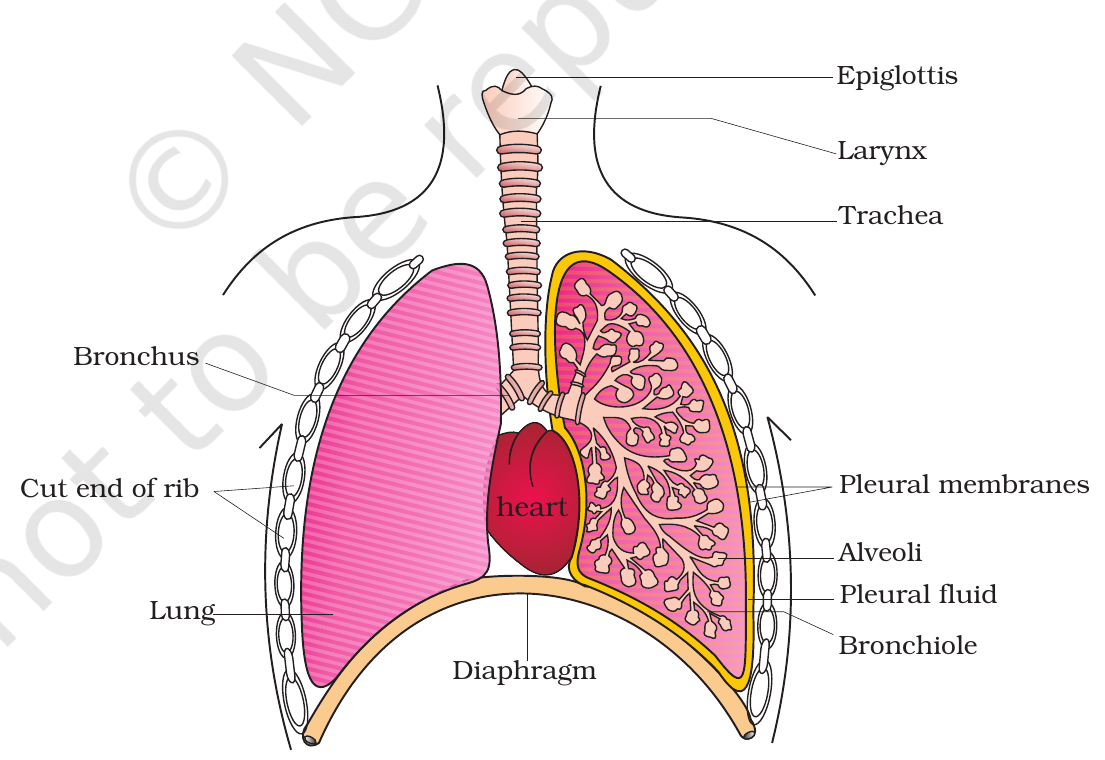
Air enters through the external nostrils and passes through the nasal passage, pharynx, larynx, trachea, bronchi, bronchioles and finally the alveoli.
- Nasal passage: fine hairs and mucus trap dust and foreign particles; the air is warmed and moistened.
- Pharynx: a common passage for food and air.
- Larynx: a cartilaginous box that helps in sound production, hence called the sound box. Its opening, the glottis, is covered by a thin elastic cartilaginous flap called the epiglottis during swallowing, so food does not enter the larynx.
- Trachea: a straight tube continuing to the mid-thoracic cavity, where it divides at the level of the 5th thoracic vertebra into a right and a left primary bronchus. The trachea, primary, secondary and tertiary bronchi, and initial bronchioles are supported by incomplete cartilaginous rings, which keep the airways open.
- Bronchi divide repeatedly into secondary and tertiary bronchi, then into bronchioles ending up in very thin terminal bronchioles. Each terminal bronchiole gives rise to a number of very thin, irregular-walled and vascularised bag-like structures called alveoli.
The conducting part runs from the external nostrils up to the terminal bronchioles. It transports the inhaled air to the alveoli, clears it of foreign particles, humidifies it and brings it to body temperature. No gas exchange happens here.
The exchange part is the alveoli and their ducts. This is the site of the actual diffusion of $O_2$ and $CO_2$ between blood and atmospheric air.
The lungs are situated in the thoracic chamber, which is anatomically an air-tight chamber. The lungs are covered by a double-layered pleura, with pleural fluid between the layers, which reduces friction on the lung surface. The thoracic chamber is formed dorsally by the vertebral column, ventrally by the sternum, laterally by the ribs and on the lower side by the dome-shaped diaphragm. The alveoli are thin-walled and have a rich network of blood capillaries.
Practice1 question
Try it now. A wrong answer costs nothing here, try again until you get it.
Which structure prevents food from entering the larynx during swallowing?
The epiglottis is a thin elastic cartilaginous flap that covers the glottis during swallowing, so food does not enter the larynx. The trachea carries air, the glottis is the opening into the trachea, and the pleura covers the lungs.
Mechanism of breathing
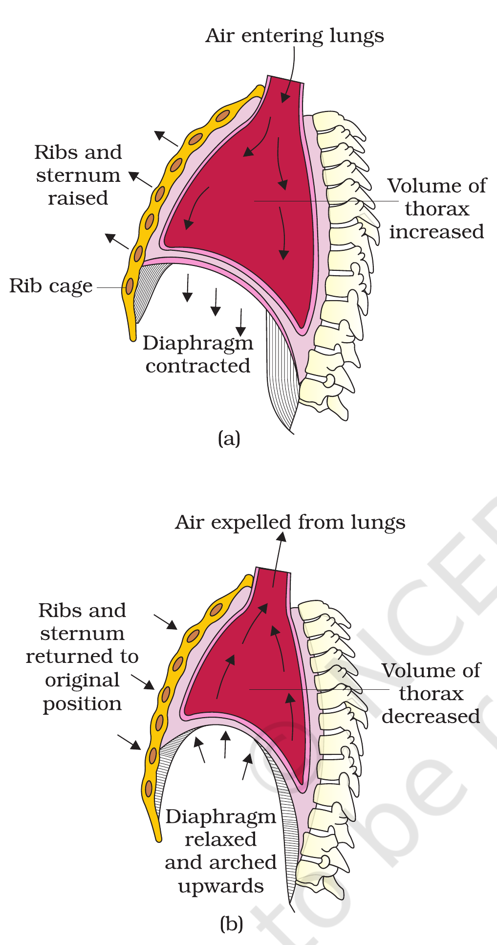
Breathing involves two stages: inspiration (air moves in) and expiration (air moves out). The movement of air into and out of the lungs is carried out by creating a pressure gradient between the lungs and the atmosphere. Inspiration can occur if the pressure within the lungs (intra-pulmonary pressure) is less than the atmospheric pressure. Similarly, expiration takes place when the intra-pulmonary pressure is higher than the atmospheric pressure.
Inspiration is initiated by the contraction of the diaphragm, which increases the volume of the thoracic chamber in the antero-posterior axis. The contraction of the external intercostal muscles lifts up the ribs and the sternum, causing an increase in the volume of the thoracic chamber in the dorso-ventral axis. The overall increase in thoracic volume causes a similar increase in pulmonary volume. An increase in pulmonary volume decreases the intra-pulmonary pressure to less than the atmospheric pressure, which forces the air from outside to move into the lungs, i.e., inspiration. On an average, a healthy human breathes 12-16 times/minute.
Relaxation of the diaphragm and the intercostal muscles returns the diaphragm and sternum to their normal positions and reduces the thoracic volume and thereby the pulmonary volume. This leads to an increase in intra-pulmonary pressure to slightly above the atmospheric pressure, causing the expulsion of air from the lungs, i.e., expiration. Normal expiration is passive; no muscles contract.
We have the ability to increase the strength of inspiration and expiration with the help of additional muscles in the abdomen.
Practice1 question
Try it now. A wrong answer costs nothing here, try again until you get it.
During inspiration, the diaphragm contracts and the thoracic volume:
Contraction of the diaphragm (it flattens and moves down) and of the external intercostal muscles increases thoracic volume, so intrapulmonary pressure falls below atmospheric pressure and air rushes in.
Respiratory volumes and capacities
Respiratory volumes are measured with a spirometer.
- Tidal Volume (TV): the volume of air inspired or expired during a normal respiration, about 500 mL. A healthy man can inspire or expire approximately 6000 to 8000 mL of air per minute.
- Inspiratory Reserve Volume (IRV): the additional volume of air a person can inspire by a forcible inspiration, about 2500 to 3000 mL.
- Expiratory Reserve Volume (ERV): the additional volume of air a person can expire by a forcible expiration, about 1000 to 1100 mL.
- Residual Volume (RV): the volume of air remaining in the lungs even after a forcible expiration, about 1100 to 1200 mL. It keeps the alveoli open.
Capacities are combinations of volumes:
- Inspiratory Capacity (IC) = TV + IRV = 3000-3500 mL: the total volume of air a person can inspire after a normal expiration.
- Expiratory Capacity (EC) = TV + ERV = 1500-1600 mL: the total volume of air a person can expire after a normal inspiration.
- Functional Residual Capacity (FRC) = ERV + RV = 2100-2300 mL: the volume of air remaining in the lungs after a normal expiration.
- Vital Capacity (VC) = ERV + TV + IRV = 4000-4600 mL: the maximum volume of air a person can breathe in after a forced expiration. This includes ERV, TV and IRV or the maximum volume of air a person can breathe out after a forced inspiration.
- Total Lung Capacity (TLC) = VC + RV = 5100-5800 mL: the total volume of air accommodated in the lungs at the end of a forced inspiration. This includes RV, ERV, TV and IRV or vital capacity + residual volume.
Practice1 question
Try it now. A wrong answer costs nothing here, try again until you get it.
A person has a tidal volume of 500 mL and an inspiratory reserve volume of 3000 mL. What is his inspiratory capacity?
Inspiratory capacity = TV + IRV = 500 + 3000 = 3500 mL.
Exchange of gases
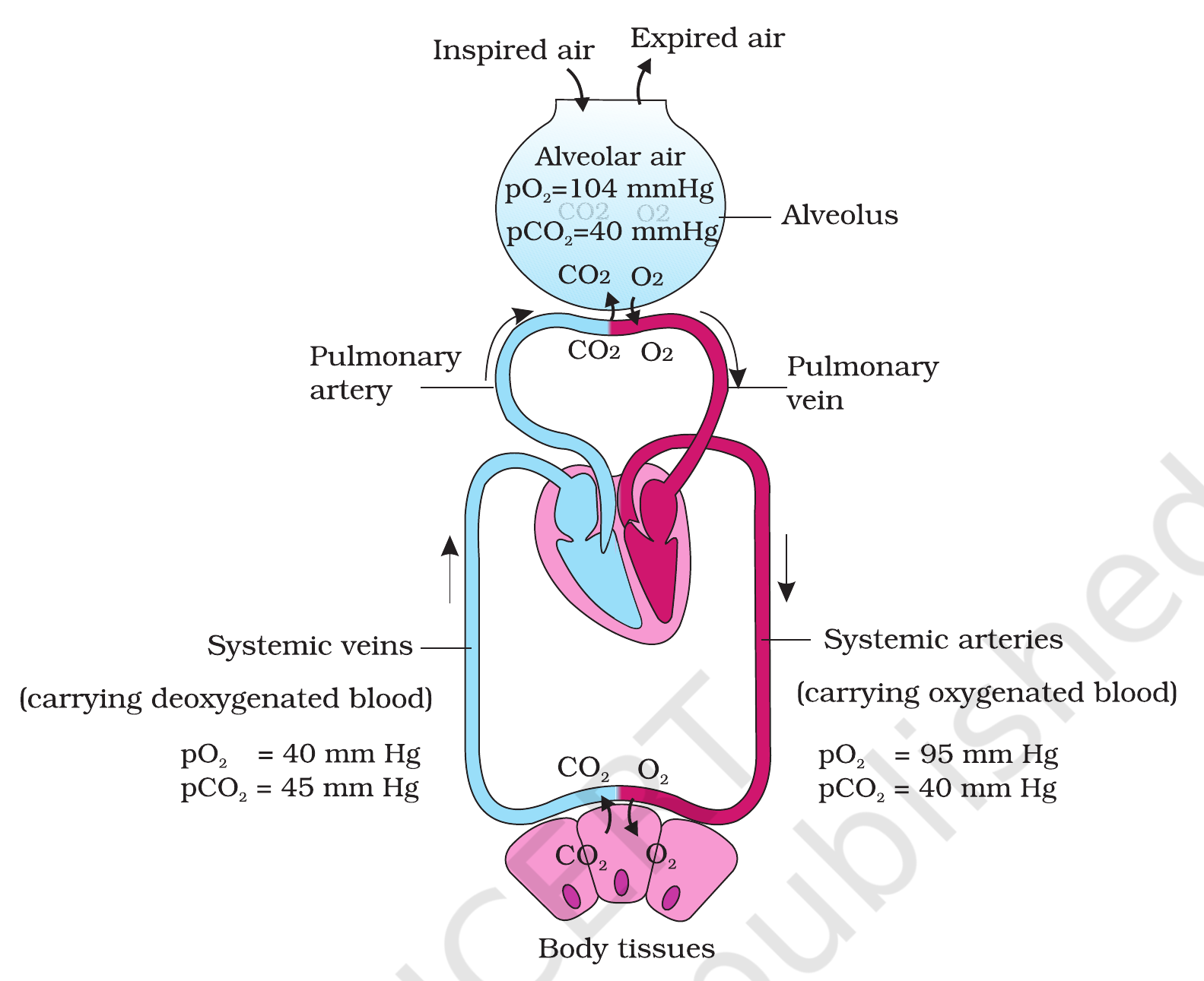
Alveoli are the primary sites of exchange of gases. Exchange of gases also occurs between blood and tissues.
Exchange happens by simple diffusion, driven by partial pressure gradients. The factors that affect the rate of diffusion are the solubility of the gases, the thickness of the membranes and the surface area available.
The partial pressures (in mm Hg) are given in NCERT Table 14.1: oxygen is 159 in atmospheric air, 104 in the alveoli, 40 in deoxygenated blood, 95 in oxygenated blood and 40 in the tissues; carbon dioxide is 0.3 in atmospheric air, 40 in the alveoli, 45 in deoxygenated blood, 40 in oxygenated blood and 45 in the tissues.
At the alveoli, the $pO_2$ of alveolar air (104) is higher than the $pO_2$ of deoxygenated blood (40), so oxygen diffuses into the blood. The $pCO_2$ of blood (45) is higher than the $pCO_2$ of alveolar air (40), so carbon dioxide diffuses out of the blood into the alveoli.
At the tissues, the $pO_2$ of oxygenated blood (95) is higher than the $pO_2$ of the tissues (40), so oxygen diffuses into the tissues. The $pCO_2$ of the tissues (45) is higher than that of the blood (40), so carbon dioxide diffuses into the blood.
The diffusion membrane is made up of three layers: the thin squamous epithelium of the alveoli, the endothelium of the alveolar capillaries, and the basement substance between them. Its total thickness is much less than a millimetre.
The solubility of carbon dioxide is 20-25 times higher than that of oxygen, so the amount of $CO_2$ that can diffuse through the diffusion membrane per unit difference in partial pressure is much higher compared to that of $O_2$.
Transport of oxygen
Blood is the medium of transport of gases. About 97 per cent of $O_2$ is transported by the red blood cells, bound to haemoglobin. The remaining 3 per cent is carried in a dissolved state through the plasma.
Haemoglobin is a red coloured iron-containing pigment in the RBCs. Each haemoglobin molecule can carry a maximum of four molecules of $O_2$, binding them reversibly to form oxyhaemoglobin: Hb + 4O2 gives Hb($O_2$)4.
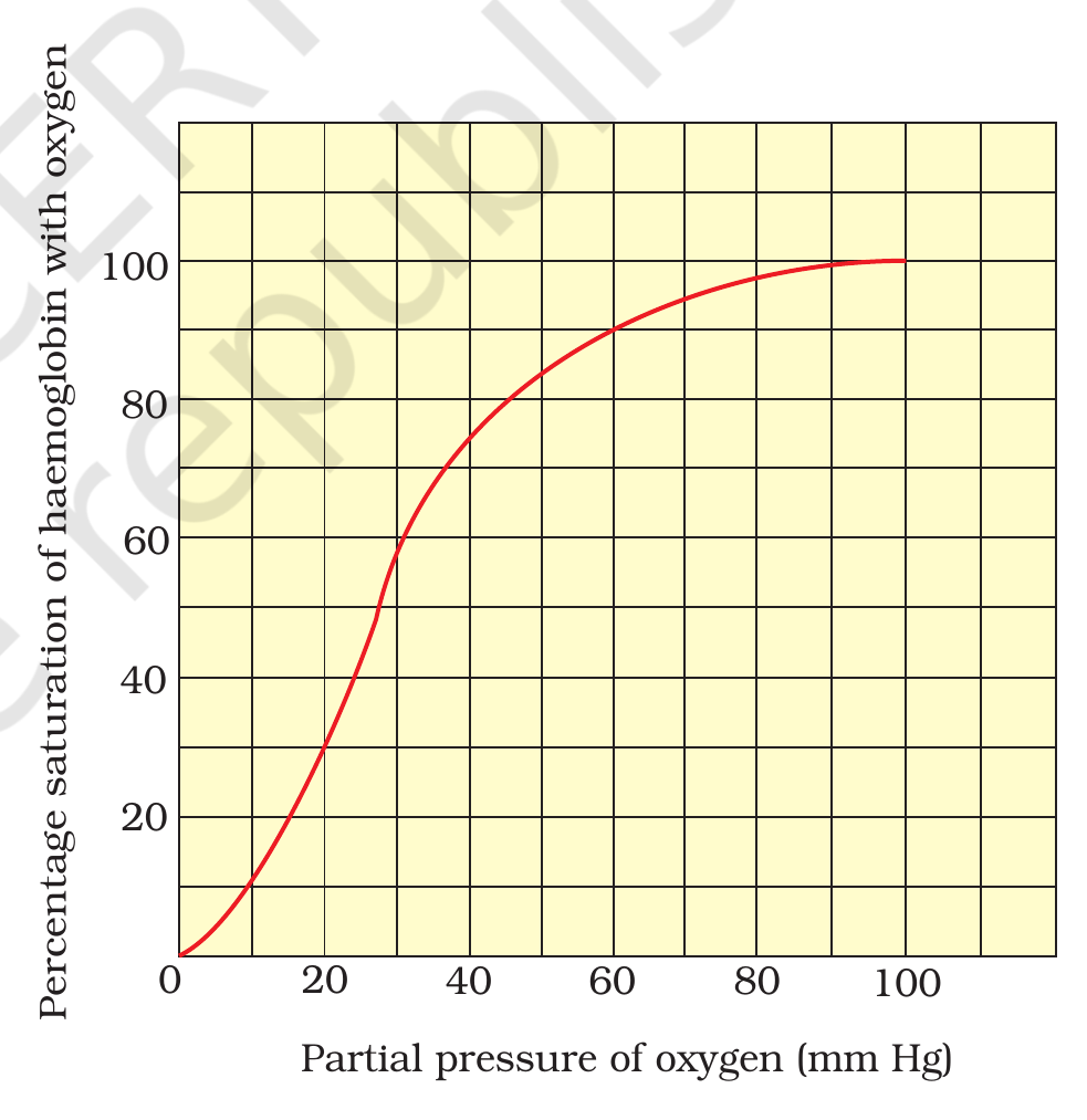
The binding of oxygen with haemoglobin is primarily related to the partial pressure of $O_2$. The partial pressure of $CO_2$, hydrogen ion concentration and temperature are the other factors that influence binding.
A sigmoid (S-shaped) curve is obtained when the percentage saturation of haemoglobin with $O_2$ is plotted against the $pO_2$. This is the oxygen dissociation curve, and it is highly useful in studying the effect of $pCO_2$, $H^+$ concentration and other factors on the binding of $O_2$ with haemoglobin.
In the alveoli, where there is high $pO_2$, low $pCO_2$, lesser $H^+$ concentration and lower temperature, the conditions are favourable for the formation of oxyhaemoglobin.
In the tissues, where there is low $pO_2$, high $pCO_2$, high $H^+$ concentration and higher temperature, the conditions are favourable for dissociation of oxygen from oxyhaemoglobin, so oxygen is released to the tissues.
Every 100 mL of oxygenated blood can deliver around 5 mL of $O_2$ to the tissues under normal physiological conditions.
Practice1 question
Try it now. A wrong answer costs nothing here, try again until you get it.
Which conditions in the alveoli favour the formation of oxyhaemoglobin?
In the alveoli, high $pO_2$, low $pCO_2$, lesser $H^+$ concentration and lower temperature favour the binding of oxygen to haemoglobin. In the tissues the opposite conditions favour dissociation.
Transport of carbon dioxide
Carbon dioxide is transported in three forms: about 70 per cent as bicarbonate ions ($HCO_3^-$), 20-25 per cent as carbaminohaemoglobin, and about 7 per cent in a dissolved state through plasma. $CO_2$ is carried by haemoglobin as carbamino-haemoglobin (about 20-25 per cent). This binding is related to the partial pressure of $CO_2$. $pO_2$ is a major factor which could affect this binding. When $pCO_2$ is high and $pO_2$ is low as in the tissues, more binding of carbon dioxide occurs whereas, when the $pCO_2$ is low and $pO_2$ is high as in the alveoli, dissociation of $CO_2$ from carbamino-haemoglobin takes place, i.e., $CO_2$ which is bound to haemoglobin from the tissues is delivered at the alveoli.
In the tissues, where $pCO_2$ is high, $CO_2$ diffuses into the blood (plasma and RBCs). In the RBCs, the enzyme carbonic anhydrase catalyses the reaction of $CO_2$ with water to form carbonic acid ($H_2CO_3$), which quickly dissociates into bicarbonate ions ($HCO_3^-$) and hydrogen ions ($H^+$).
Carbonic anhydrase is present in high concentration in the RBCs and in small amounts in the plasma.
At the alveoli, where $pCO_2$ is low, the reaction proceeds in the opposite direction, leading to the formation of $CO_2$ and $H_2O$. Thus, $CO_2$ trapped as bicarbonate at the tissue level and transported to the alveoli is released out as $CO_2$ and exhaled.
Every 100 mL of deoxygenated blood delivers approximately 4 mL of $CO_2$ to the alveoli.
Regulation of respiration
The respiratory rhythm centre is situated in the medulla region of the brain and is primarily responsible for the regulation of respiration.
A pneumotaxic centre is situated in the pons region of the brain. It can moderate the functions of the respiratory rhythm centre: a neural signal from this centre can reduce the duration of inspiration and thereby alter the respiratory rate.
A chemosensitive area is situated adjacent to the rhythm centre and is highly sensitive to $CO_2$ and hydrogen ions. An increase in these substances activates this area, which in turn signals the rhythm centre to make the necessary adjustments in the respiratory process.
Receptors associated with the aortic arch and the carotid artery also recognise changes in $CO_2$ and $H^+$ concentration and send the necessary signals to the rhythm centre for remedial actions.
The role of oxygen in the regulation of respiratory rhythm is quite insignificant.
Practice1 question
Try it now. A wrong answer costs nothing here, try again until you get it.
The pneumotaxic centre, which can reduce the duration of inspiration and alter the respiratory rate, is located in the:
The pneumotaxic centre is situated in the pons of the brain. The respiratory rhythm centre is in the medulla; the chemosensitive area lies adjacent to it.
Disorders of respiratory system
Asthma is a difficulty in breathing that causes wheezing. It is due to inflammation of the bronchi and bronchioles.
Emphysema is a chronic disorder in which the alveolar walls are damaged and the respiratory surface is decreased. One of the major causes of this is cigarette smoking.
Occupational respiratory disorders occur in certain industries, especially those involving grinding or stone-breaking, where so much dust is produced that the defence mechanism of the body cannot fully cope with the situation. Long exposure can give rise to inflammation leading to fibrosis (proliferation of fibrous tissues) of the lungs. This results in serious lung damage. Workers in such industries should wear protective masks.
Table 1 - Respiratory organs across animals
| Animal | Respiratory organ |
|---|---|
| Sponges, coelenterates, flatworms | Entire body surface (simple diffusion) |
| Earthworm | Moist cuticle |
| Insects | Tracheal tubes |
| Aquatic arthropods and molluscs | Gills |
| Fishes | Gills |
| Amphibians (frog) | Lungs and moist skin |
| Reptiles, birds, mammals | Lungs |
Source: NCERT XI, Ch 14, 14.1.
Table 2 - Respiratory volumes and capacities
| Volume or capacity | Definition | Value |
|---|---|---|
| Tidal Volume (TV) | Air inspired or expired during normal respiration | 500 mL |
| Inspiratory Reserve Volume (IRV) | Additional air inspired by forcible inspiration | 2500-3000 mL |
| Expiratory Reserve Volume (ERV) | Additional air expired by forcible expiration | 1000-1100 mL |
| Residual Volume (RV) | Air remaining in lungs after forcible expiration | 1100-1200 mL |
| Inspiratory Capacity (IC = TV + IRV) | Total air inspired after a normal expiration | 3000-3500 mL |
| Expiratory Capacity (EC = TV + ERV) | Total air expired after a normal inspiration | 1500-1600 mL |
| Functional Residual Capacity (FRC = ERV + RV) | Air remaining after a normal expiration | 2100-2300 mL |
| Vital Capacity (VC = ERV + TV + IRV) | Maximum air breathed in after forced expiration, or breathed out after forced inspiration | 4000-4600 mL |
| Total Lung Capacity (TLC = VC + RV) | Total air in lungs after maximum inspiration | 5100-5800 mL |
Source: NCERT XI, Ch 14, 14.2.1.
Table 3 - Partial pressures of gases (NCERT Table 14.1)
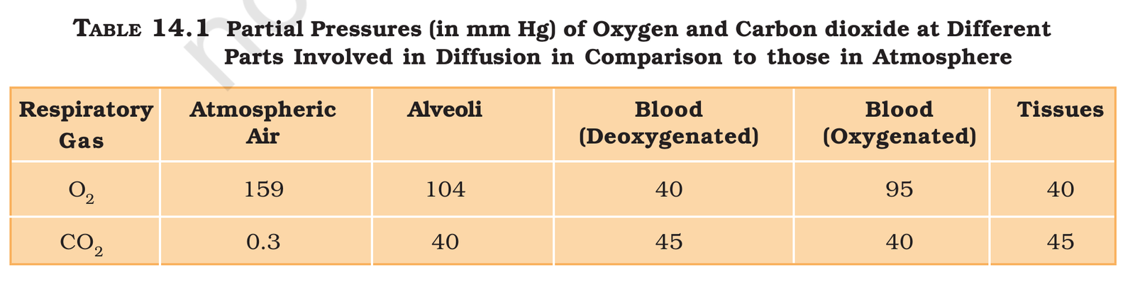
| Respiratory gas | Atmospheric air | Alveoli | Blood (deoxygenated) | Blood (oxygenated) | Tissues |
|---|---|---|---|---|---|
| Oxygen | 159 | 104 | 40 | 95 | 40 |
| Carbon dioxide | 0.3 | 40 | 45 | 40 | 45 |
All values in mm Hg. Source: NCERT XI, Ch 14, Table 14.1.
Table 4 - Conditions for oxyhaemoglobin formation and dissociation
| Condition | In the alveoli (formation favoured) | In the tissues (dissociation favoured) |
|---|---|---|
| $pO_2$ | High | Low |
| $pCO_2$ | Low | High |
| $H^+$ concentration | Lesser | Higher |
| Temperature | Lower | Higher |
Source: NCERT XI, Ch 14, 14.4.1.
Trap pairs: the confusions NEET tests
- Inspiration vs expiration: inspiration is active (diaphragm and external intercostal muscles contract); normal expiration is passive (the muscles relax). Only forced expiration uses the abdominal muscles.
- Conducting part vs exchange part: the conducting part (external nostrils to terminal bronchioles) only transports, clears, humidifies and warms the air; exchange happens only in the alveoli and their ducts.
- IRV vs ERV vs RV: IRV is extra air you can breathe IN by forceful inspiration (2500-3000 mL); ERV is extra air you can breathe OUT by forceful expiration (1000-1100 mL); RV is the air that stays even after forceful expiration (1100-1200 mL) and keeps the alveoli from collapsing.
- $O_2$ vs $CO_2$ transport: 97 per cent of $O_2$ rides on haemoglobin; 70 per cent of $CO_2$ travels as bicarbonate ions. The dominant form is different for each gas.
- Asthma vs emphysema: asthma is inflammation of bronchi and bronchioles, causing wheezing; emphysema is damage to alveolar walls, decreasing the respiratory surface, mostly from cigarette smoking.
- $pO_2$ at alveoli vs tissues: alveoli 104 mm Hg, tissues 40 mm Hg. The gradient drives $O_2$ into the blood at the alveoli and out of the blood into the tissues.
- Chemosensitive area vs pneumotaxic centre: the chemosensitive area (adjacent to the rhythm centre in the medulla) responds to $CO_2$ and $H^+$; the pneumotaxic centre (in the pons) limits the duration of inspiration and alters the respiratory rate.
- Residual volume keeps alveoli open: the lungs do not collapse even after forceful expiration because of the residual volume (NEET 2017).
Trap Boxes
Expiration is an active process?
Wrong idea: Expiration, like inspiration, needs muscles to contract.
Normal expiration is passive: the diaphragm and external intercostal muscles relax, thoracic volume falls, intrapulmonary pressure rises and air is pushed out. Only forced expiration uses the abdominal muscles.
NCERT XI, Ch 14, 14.2
Gas exchange happens in the bronchi?
Wrong idea: Oxygen and carbon dioxide are exchanged all along the airways, including the bronchi.
Exchange happens only in the alveoli and their ducts. The conducting part (nostrils to terminal bronchioles) only transports, clears, humidifies and warms the air.
NCERT XI, Ch 14, 14.1
Most carbon dioxide travels on haemoglobin?
Wrong idea: Like oxygen, most carbon dioxide is carried bound to haemoglobin.
Only 20-25 per cent of $CO_2$ is carried as carbaminohaemoglobin. About 70 per cent travels as bicarbonate ions and 7 per cent dissolved in plasma.
NCERT XI, Ch 14, 14.4.2
Asthma and emphysema are the same problem?
Wrong idea: Asthma and emphysema both mean the alveoli are damaged.
Asthma is inflammation of bronchi and bronchioles causing wheezing. Emphysema is damage to alveolar walls, decreasing the respiratory surface, mostly from cigarette smoking.
NCERT XI, Ch 14, 14.6
The lungs collapse after forceful expiration?
Wrong idea: After a forceful expiration the lungs collapse because all the air is out.
The residual volume (1100-1200 mL) stays in the lungs even after forceful expiration and keeps the alveoli open.
NCERT XI, Ch 14, 14.2.1
Chapter test
5 NEET PYQs. +4 for a correct answer, -1 for a wrong answer, exactly like NEET. Finish the test to earn a medal.
Q1: NEET (UG) 2021, Code M1, Q179
Select the favourable conditions required for the formation of oxyhaemoglobin at the alveoli.
In the alveoli, high $pO_2$, low $pCO_2$, lesser $H^+$ concentration and lower temperature favour the formation of oxyhaemoglobin. Only option (1) lists all four alveolar conditions; the other options contain tissue conditions that favour dissociation.
Q2: NEET (UG) 2021, Code M1, Q183
The partial pressures (in mm Hg) of oxygen ($O_2$) and carbon dioxide ($CO_2$) at alveoli (the site of diffusion) are:
At the alveoli the partial pressure of oxygen is 104 mm Hg and that of carbon dioxide is 40 mm Hg (NCERT Table 14.1). Oxygen diffuses from the alveoli into the blood because alveolar $pO_2$ (104) is higher than the $pO_2$ of deoxygenated blood (40).
Q3: NEET (UG) 2019, Code S2, Q27
Tidal volume and expiratory reserve volume of an athlete is 500 mL and 1000 mL respectively. What will be his expiratory capacity if the residual volume is 1200 mL?
Expiratory capacity = TV + ERV = 500 + 1000 = 1500 mL. The residual volume (1200 mL) is not part of expiratory capacity; it is part of functional residual capacity and total lung capacity.
Q4: NEET (UG) 2018, Code AA, Q136
Which of the following options correctly represents the lung conditions in asthma and emphysema, respectively?
Asthma is difficulty in breathing causing wheezing due to inflammation of bronchi and bronchioles. Emphysema is a chronic disorder in which alveolar walls are damaged and the respiratory surface is decreased.
Q5: NEET (UG) 2017, Code A, Q148
Lungs are made up of air-filled sacs, the alveoli. They do not collapse even after forceful expiration, because of:
The residual volume (1100-1200 mL) remains in the lungs even after forceful expiration, so the alveoli stay open and do not collapse.
Score: 0 · Answered 0 of 5
Flashcards
Tap a card to flip it over and see the answer. Tap it again to flip it back.
What is the tidal volume in a normal healthy man?
Tap to flip500 mL.
Tap to flip backWhat are the values of IRV, ERV and RV?
Tap to flipIRV 2500-3000 mL; ERV 1000-1100 mL; RV 1100-1200 mL.
Tap to flip backPut these in the order air travels through them: larynx, alveoli, pharynx, trachea, bronchi, bronchioles.
Tap to flipPharynx, larynx, trachea, bronchi, bronchioles, alveoli.
Tap to flip backWhat is the partial pressure of oxygen in the alveoli and in the tissues?
Tap to flipAlveoli 104 mm Hg; tissues 40 mm Hg.
Tap to flip backWhat percentage of oxygen is transported by haemoglobin, and what percentage of carbon dioxide as bicarbonate?
Tap to flip97 per cent of $O_2$ is transported by RBCs (haemoglobin); 70 per cent of $CO_2$ is transported as bicarbonate.
Tap to flip backWhich part of the respiratory system is the conducting part, and what does it NOT do?
Tap to flipExternal nostrils to terminal bronchioles; it does not exchange gases.
Tap to flip backName the three forms in which carbon dioxide is transported in blood.
Tap to flipAs bicarbonate (70 per cent), as carbaminohaemoglobin (20-25 per cent), dissolved in plasma (7 per cent).
Tap to flip backPut in order: $CO_2$ diffuses into blood at the tissues, carbonic anhydrase forms carbonic acid, the acid splits into bicarbonate and $H^+$, the reaction reverses at the alveoli and $CO_2$ is released.
Tap to flipAt the tissues, where $pCO_2$ is high, $CO_2$ diffuses into the blood and forms $HCO_3^-$ and $H^+$; at the alveoli, where $pCO_2$ is low, the reaction runs in reverse and $CO_2$ is released out. RBCs contain a very high concentration of carbonic anhydrase; minute quantities are present in the plasma too.
Tap to flip backWhere is the respiratory rhythm centre, and where is the pneumotaxic centre?
Tap to flipRespiratory rhythm centre in the medulla; pneumotaxic centre in the pons.
Tap to flip backWhich gas is 20-25 times more soluble, and why does it matter?
Tap to flipCarbon dioxide is 20-25 times more soluble than oxygen, so it diffuses across the diffusion membrane much faster per unit difference in partial pressure.
Tap to flip back